
Human 4-1BB/TNFRSF9/CD137 Antibody
R&D Systems, part of Bio-Techne | Catalog # AF838


Scientific images may be resized, cropped, or adjusted for brightness or contrast for presentation purposes. These updates are limited to presentation and do not affect the underlying scientific interpretation of the data. Where available, additional source data may be provided upon request.
Conjugate
Catalog #
Key Product Details
Validated by
Biological Validation
Species Reactivity
Validated:
Human
Cited:
Human
Applications
Validated:
Immunohistochemistry, Western Blot, ELISA Capture (Matched Antibody Pair), Flow Cytometry, Immunocytochemistry, Simple Western, Agonist Activity, CyTOF-ready
Cited:
Immunohistochemistry-Frozen, Flow Cytometry, Functional Assay
Label
Unconjugated
Antibody Source
Polyclonal Goat IgG
Product Specifications
Immunogen
Mouse myeloma cell line NS0-derived recombinant human 4‑1BB/TNFRSF9/CD137
Leu24-Gln186
Accession # Q07011
Leu24-Gln186
Accession # Q07011
Specificity
Detects 4‑1BB/TNFRSF9/CD137 in direct ELISAs and Western blots.
Clonality
Polyclonal
Host
Goat
Isotype
IgG
Endotoxin Level
<0.10 EU per 1 μg of the antibody by the LAL method.
Scientific Data Images for Human 4-1BB/TNFRSF9/CD137 Antibody
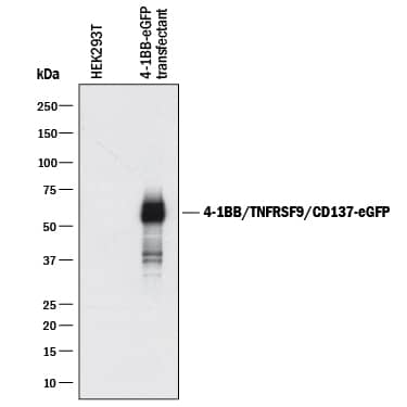
Detection of Human 4‑1BB/TNFRSF9/ CD137 by Western Blot.
Western blot shows lysates of HEK293T human embryonic kidney cell line either mock transfected or transfected with human 4-1BB/TNFRSF9/ CD137-eGFP Fusion. PVDF membrane was probed with 0.5 µg/mL of Goat Anti-Human 4-1BB/ TNFRSF9/CD137 Antigen Affinity-purified Polyclonal Antibody (Catalog # AF838) followed by HRP-conjugated Anti-Goat IgG Secondary Antibody ([catalogNumber:HAF017]]). A specific band was detected for 4-1BB/ TNFRSF9/CD137-eGFP Fusion at approximately 50-65 kDa (as indicated). This experiment was conducted under reducing conditions and using Immunoblot Buffer Group 1.
4‑1BB/TNFRSF9/CD137 in Human Tonsil.
4-1BB/TNFRSF9/CD137 was detected in immersion fixed paraffin-embedded sections of human tonsil using Goat Anti-Human 4-1BB/TNFRSF9/CD137 Antigen Affinity-purified Polyclonal Antibody (Catalog # AF838) at 10 µg/mL overnight at 4 °C. Tissue was stained using the Anti-Goat HRP-DAB Cell & Tissue Staining Kit (brown; CTS008) and counterstained with hematoxylin (blue). Specific staining was localized to lymphocytes. View our protocol for Chromogenic IHC Staining of Paraffin-embedded Tissue Sections.
Detection of 4‑1BB/TNFRSF9/CD137 in PHA-treated Human T Cells by Flow Cytometry.
Human T cells were treated for 48 hours with 5 µg/mL PHA then stained with Goat Anti-Human 4-1BB/TNFRSF9/ CD137 Antigen Affinity-purified Polyclonal Antibody (Catalog # AF838) followed by Allophycocyanin-conjugated Anti-Goat IgG Secondary Antibody (F0108) and PE-conjugated anti-human CD3 (Catalog # FAB100P). Quad marker was set based on control antibody (AB-108-C) staining.


Applications for Human 4-1BB/TNFRSF9/CD137 Antibody
Application
Recommended Usage
Agonist Activity
Measured by its ability to co-stimulate IFN-gamma secretion by human T cells in the presence of anti-CD3.
The ED50 for this effect is typically ≤ 20 μg/mL.
The ED50 for this effect is typically ≤ 20 μg/mL.
CyTOF-ready
Ready to be labeled using established conjugation methods. No BSA or other carrier proteins that could interfere with conjugation.
Flow Cytometry
0.25 µg/106 cells
Sample: Human T cells treated with PHA
Sample: Human T cells treated with PHA
Immunocytochemistry
5-15 µg/mL
Sample: Immersion fixed human peripheral blood mononuclear cells
Sample: Immersion fixed human peripheral blood mononuclear cells
Immunohistochemistry
5-25 µg/mL
Sample: Immersion fixed paraffin-embedded sections of human tonsil
Sample: Immersion fixed paraffin-embedded sections of human tonsil
Simple Western
20 µg/mL
Sample: HDLM-2 human Hodgkin's lymphoma cells
Sample: HDLM-2 human Hodgkin's lymphoma cells
Western Blot
0.5 µg/mL
Sample: HEK293T human embryonic kidney cell line transfected with human 4-1BB/TNFRSF9/CD137-eGFP Fusion
Sample: HEK293T human embryonic kidney cell line transfected with human 4-1BB/TNFRSF9/CD137-eGFP Fusion
Human 4-1BB/TNFRSF9/CD137 Sandwich Immunoassay
Please Note: Optimal dilutions of this antibody should be experimentally determined.
Reviewed Applications
Read 1 review rated 5 using AF838 in the following applications:
Formulation, Preparation, and Storage
Purification
Antigen Affinity-purified
Reconstitution
Reconstitute at 0.2 mg/mL in sterile PBS. For liquid material, refer to CoA for concentration.
Formulation
Lyophilized from a 0.2 μm filtered solution in PBS with Trehalose. See Certificate of Analysis for details.
*Small pack size (-SP) is supplied either lyophilized or as a 0.2 µm filtered solution in PBS.
*Small pack size (-SP) is supplied either lyophilized or as a 0.2 µm filtered solution in PBS.
Shipping
Lyophilized product is shipped at ambient temperature. Liquid small pack size (-SP) is shipped with polar packs. Upon receipt, store immediately at the temperature recommended below.
Stability & Storage
Use a manual defrost freezer and avoid repeated freeze-thaw cycles.
- 12 months from date of receipt, -20 to -70 °C as supplied.
- 1 month, 2 to 8 °C under sterile conditions after reconstitution.
- 6 months, -20 to -70 °C under sterile conditions after reconstitution.
Background: 4-1BB/TNFRSF9/CD137
4-1BB is an inducible T cell surface protein belonging to the TNF receptor superfamily. It is alternatively known as TNFRSF9, CD137, and ILA. The 255 amino acid human 4-1BB is a type I transmembrane protein having in its extracellular domain four of the cysteine-rich motifs that are characteristic of the TNF receptor superfamily. The 30 kDa glycoprotein exists both as a monomer and as a dimer on T cells. The human and mouse proteins share 60% amino acid identity. 4-1BB is absent from naive T cells, but it is upregulated and continually expressed following T cell activation. The natural ligand, 4-1BBL, is a member of the TNF superfamily and is expressed on activated antigen presenting cells including dendritic cells, macrophages, and B cells. Cross-linking of 4-1BB by 4-1BBL or by agonistic antibodies transmits a potent co-stimulatory signal that enhances the effect of other activating signals such as PHA or anti-CD3 antibodies. 4-1BB signals through the
TFAF2‑NIK pathway resulting in activation of NF-kappa B and ultimately promoting proliferation and survival of T cells.
References
- Vinay, D. and B. Kwon (1998) Semin. Immunol. 10:481.
- Sica, G. and L. Chen (2000) Adv. Exp. Med. Biol. 465:355.
Alternate Names
41BB, CD137, ILA, TNFRSF9
Gene Symbol
TNFRSF9
UniProt
Additional 4-1BB/TNFRSF9/CD137 Products
Product Documents for Human 4-1BB/TNFRSF9/CD137 Antibody
Product Specific Notices for Human 4-1BB/TNFRSF9/CD137 Antibody
For research use only
Loading...
Loading...
Loading...
Loading...
Loading...
Loading...