
Human TRAIL R2/TNFRSF10B PE-conjugated Antibody
R&D Systems, part of Bio-Techne | Catalog # FAB6311P


Scientific images may be resized, cropped, or adjusted for brightness or contrast for presentation purposes. These updates are limited to presentation and do not affect the underlying scientific interpretation of the data. Where available, additional source data may be provided upon request.
Conjugate
Catalog #
Key Product Details
Species Reactivity
Validated:
Human
Cited:
Human, Mouse, Rat
Applications
Validated:
Flow Cytometry
Cited:
Western Blot, Flow Cytometry
Label
Phycoerythrin (Excitation = 488 nm, Emission = 565-605 nm)
Antibody Source
Monoclonal Mouse IgG2B Clone # 71908
Product Specifications
Immunogen
Mouse myeloma cell line NS0-derived recombinant human TRAIL R2/TNFRSF10B
Ile56-Glu182
Accession # O14763
Ile56-Glu182
Accession # O14763
Specificity
Detects human TRAIL R2 in direct ELISAs and Western blots. In direct ELISAs, does not cross-react with recombinant human (rh) TRAIL R1, rhTRAIL R3, rhTRAIL R4, or recombinant mouse TRAIL R2.
Clonality
Monoclonal
Host
Mouse
Isotype
IgG2B
Scientific Data Images for Human TRAIL R2/TNFRSF10B PE-conjugated Antibody
MDA‑MB‑231 Human Cell Line by Flow Cytometry.
MDA-MB-231 human breast cancer cell line was stained with Mouse Anti-Human TRAIL R2/TNFRSF10B PE-conjugated Monoclonal Antibody (Catalog # FAB6311P, filled histogram) or isotype control antibody (Catalog # IC0041P, open histogram). View our protocol for Staining Membrane-associated Proteins.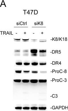
Detection of TRAIL R2/TNFRSF10B by Western Blot
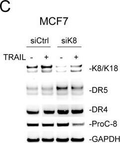
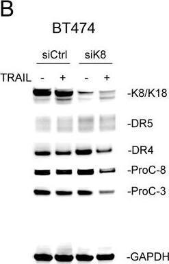
Knockdown of K8 enhances TRAIL induced apoptosis(A-C) Cells were transfected with a control siRNA or siRNA specific to KRT8 transcript for 72 hours, followed by TRAIL stimulation (100 ng/ml [T47D and BT474] or 150 ng/ml [MCF7]) for 24 hours. The resultant cells were analyzed by immunoblotting using antibodies specific to K8/K18, DR4, DR5, caspase-3, caspase-8, and glyceraldehyde 3-phosphate dehydrogenase (GAPDH). Caspase activation was identified by decrease in pro-enzyme form (ProC-8 and ProC-3). (D-F) Cells were treated as above and analyzed by flow cytometry after staining with Annexin-V-FITC and propidium iodide (PI). Image collected and cropped by CiteAb from the following open publication (https://pubmed.ncbi.nlm.nih.gov/29796187), licensed under a CC-BY license. Not internally tested by R&D Systems.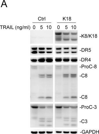
Detection of TRAIL R2/TNFRSF10B by Western Blot
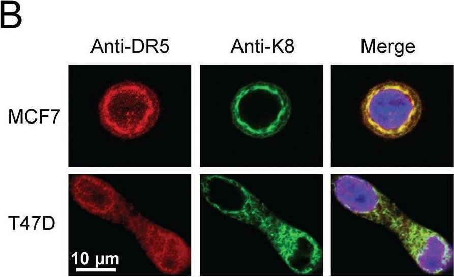
Keratin 8/18 overexpression downregulates total DR5 protein levels(A) MDA-MB-231 cells were transfected with a control plasmid or plasmid encoding human keratin 18 for 24 hours, followed by TRAIL stimulation (0, 5, 10 ng/ml) for 3 hours. The resultant cells were analyzed by immunoblotting using antibodies against K8/K18, DR5, DR4, caspase-8, caspase-3, and glyceraldehyde 3-phosphate dehydrogenase (GAPDH). Caspase activation is indicated by a decrease in pro-enzyme form (ProC-8 and ProC-3) and by the simultaneous appearance of cleaved fragments. Results are from non-adjoining lanes from the same gel. (B) Relative total DR5 and DR4 protein levels were quantified using densitometry analysis of immunoblots in A and were normalized to GAPDH loading controls. P-values were determined using a student’s t-test. Image collected and cropped by CiteAb from the following open publication (https://pubmed.ncbi.nlm.nih.gov/29796187), licensed under a CC-BY license. Not internally tested by R&D Systems.
Applications for Human TRAIL R2/TNFRSF10B PE-conjugated Antibody
Application
Recommended Usage
Flow Cytometry
10 µL/106 cells
Sample: MDA‑MB‑231 human breast cancer cell line
Sample: MDA‑MB‑231 human breast cancer cell line
Reviewed Applications
Read 1 review rated 4 using FAB6311P in the following applications:
Spectra Viewer
Plan Your Experiments
Use our spectra viewer to interactively plan your experiments, assessing multiplexing options. View the excitation and emission spectra for our fluorescent dye range and other commonly used dyes.
Spectra Viewer
Formulation, Preparation, and Storage
Purification
Protein A or G purified from hybridoma culture supernatant
Formulation
Supplied in a saline solution containing BSA and Sodium Azide.
Shipping
The product is shipped with polar packs. Upon receipt, store it immediately at the temperature recommended below.
Stability & Storage
Protect from light. Do not freeze.
- 12 months from date of receipt, 2 to 8 °C as supplied.
Background: TRAIL R2/TNFRSF10B
Human TRAIL R2, also known as DR5 and TRICK 2, is a type 1, TNF R family, transmembrane protein which is a receptor for TRAIL (APO2 Ligand). In the new TNF superfamily nomenclature, TRAIL R2 is referred to as TNFRSF10B. TRAIL R2 cDNA encodes a 440 amino acid residue precursor protein containing extracellular cysteine-rich domains, a transmembrane domain and a cytoplasmic death domain. Among TNF receptor family proteins, TRAIL R2 is most closely related to TRAIL R1/DR4, sharing 55% amino acid sequence identity. Binding of trimeric TRAIL to TRAIL R2 induces apoptosis. The induction of apoptosis likely requires oligomerization of the receptor. The human TRAIL R2/Fc chimera neutralizes the ability of TRAIL to induce apoptosis. Besides TRAIL R2, an additional TRAIL R1/DR4, which tranduces apoptosis signaling, and two TRAIL decoy receptors, which antagonize TRAIL-induced apoptosis, have been reported.
Long Name
TRAIL Receptor 2
Alternate Names
CD262, DR5, TNFRSF10B, TRAILR2
Gene Symbol
TNFRSF10B
UniProt
Additional TRAIL R2/TNFRSF10B Products
Product Documents for Human TRAIL R2/TNFRSF10B PE-conjugated Antibody
Product Specific Notices for Human TRAIL R2/TNFRSF10B PE-conjugated Antibody
For research use only
Loading...
Loading...
Loading...
Loading...
Loading...