Human PD‑1 Antibody
Catalog # AF1086 | R&D Systems, Inc. a Bio-Techne Brand
Key Product Details
Species Reactivity
Applications
Label
Antibody Source
Customers Also Viewed
Product Summary for Human PD‑1 Antibody
Immunogen
Leu25-Gln167
Accession # Q8IX89
Specificity
Clonality
Host
Isotype
Scientific Data Images for Human PD‑1 Antibody
Detection of Human PD‑1 by Western Blot.
Western blot shows lysate of human thymus tissue. PVDF membrane was probed with 2 µg/mL of Goat Anti-Human PD-1 Antigen Affinity-purified Polyclonal Antibody (Catalog # AF1086) followed by HRP-conjugated Anti-Goat IgG Secondary Antibody (Catalog # HAF017). Specific bands were detected for PD-1 at approximately 40-50 kDa (as indicated). This experiment was conducted under reducing conditions and using Immunoblot Buffer Group 1.Detection of Human PD‑1 by Western Blot.
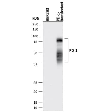
Western blot shows lysates of HEK293 human embryonic kidney cell line either mock transfected or transfected with human PD-1. PVDF membrane was probed with 0.5 µg/mL of Goat Anti-Human PD-1 Antigen Affinity-purified Polyclonal Antibody (Catalog # AF1086) followed by HRP-conjugated Anti-Goat IgG Secondary Antibody (Catalog # HAF017). Specific bands were detected for PD-1 at approximately 40-80 kDa (as indicated). This experiment was conducted under reducing conditions and using Immunoblot Buffer Group 1.Detection of PD‑1 in Human PBMCs treated with PHA by Flow Cytometry.
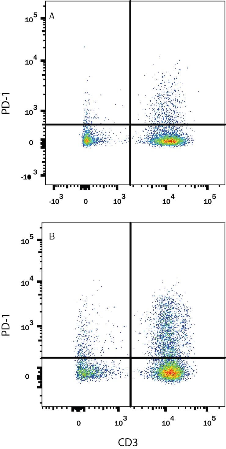
Human peripheral blood mononuclear cells (PBMCs) either (A) untreated or (B) treated with 5 µg/mL PHA overnight were stained with Goat Anti-Human PD-1 Antigen Affinity-purified Polyclonal Antibody (Catalog # AF1086) followed by Phycoerythrin-conjugated Anti-Goat IgG Secondary Antibody (Catalog # F0107) and Mouse Anti-Human CD3e APC-conjugated Monoclonal Antibody (Catalog # FAB100A). Quadrant markers were set based on control antibody staining (Catalog # F0107). View our protocol for Staining Membrane-associated Proteins.Applications for Human PD‑1 Antibody
Blockade of Receptor-ligand Interaction
CyTOF-ready
Flow Cytometry
Sample: Human peripheral blood mononuclear cells (PBMCs) treated with PHA
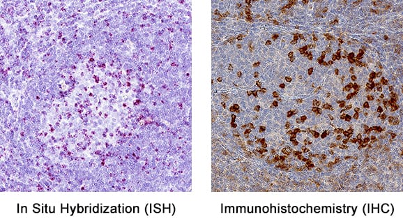
Immunohistochemistry
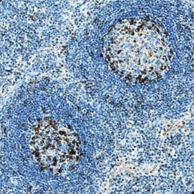
Sample: Immersion fixed paraffin-embedded sections of human lymph node
Western Blot
Sample: Human thymus tissue and HEK293 human embryonic kidney cell line transfected with human PD-1
Human PD-1 Sandwich Immunoassay
Reviewed Applications
Read 6 reviews rated 4.5 using AF1086 in the following applications:
Formulation, Preparation and Storage
Purification
Endotoxin Level
Reconstitution
Formulation
Shipping
Stability & Storage
- 12 months from date of receipt, -20 to -70 °C as supplied.
- 1 month, 2 to 8 °C under sterile conditions after reconstitution.
- 6 months, -20 to -70 °C under sterile conditions after reconstitution.
Background: PD-1
Programmed Death-1 (PD-1) is a type I transmembrane protein belonging to the CD28/CTLA-4 family of immunoreceptors that mediate signals for regulating immune responses (1). Members of the CD28/CTLA-4 family have been shown to either promote T cell activation (CD28 and ICOS) or down-regulate T cell activation (CTLA-4 and PD-1) (2). PD-1 is expressed on activated T cells, B cells, myeloid cells, and on a subset of thymocytes. In vitro, ligation of PD-1 inhibits TCR-mediated T-cell proliferation and production of IL-1, IL-4, IL-10, and IFN-gamma. In addition, PD-1 ligation also inhibits BCR mediated signaling. PD-1 deficient mice have a defect in peripheral tolerance and spontaneously develop autoimmune diseases (2, 3).
Two B7 family proteins, PD-L1 (also called B7-H1) and PD-L2 (also known as B7-DC), have been identified as PD-1 ligands. Unlike other B7 family proteins, both
PD‑L1 and PD-L2 are expressed in a wide variety of normal tissues including heart, placenta, and activated spleens (4). The wide expression of PD-L1 and PD-L2 and the inhibitor effects on PD-1 ligation indicate that PD-1 might be involved in the regulation of peripheral tolerance and may help prevent autoimmune diseases (2).
The human PD-1 gene encodes a 288 amino acid (aa) protein with a putative 20 aa signal peptide, a 148 aa extracellular region with one immunoglobulin-like V-type domain, a 24 aa transmembrane domain, and a 95 aa cytoplasmic region. The cytoplasmic tail contains two tyrosine residues that form the immuno-receptor
tyrosine-based inhibitory motif (ITIM) and immunoreceptor tyrosine-based switch motif (ITSM) that are important in mediating PD-1 signaling. Mouse and human PD-1 share approximately 60% aa sequence identity (4).
References
- Ishida, Y. et al. (1992) EMBO J. 11:3887.
- Nishimura, H. and T. Honjo (2001) Trends in Immunol. 22:265.
- Latchman, Y. et al. (2001) Nature Immun. 2:261.
- Carreno, B.M. and M. Collins (2002) Annu. Rev. Immunol. 20:29.
Long Name
Alternate Names
Entrez Gene IDs
Gene Symbol
UniProt
Product Documents for Human PD‑1 Antibody
Product Specific Notices for Human PD‑1 Antibody
For research use only
Citations for Human PD‑1 Antibody (34)
Citations are publications that use Bio-Techne products. Selected citations for Human PD‑1 Antibody include:
-
Species: Human
Sample Types: Protein
Applications: Western BlotH�nze et al. (2023-05-03), Modulation of immune checkpoint regulators in interferon ? induced urothelial carcinoma and activated T-lymphocyte cells by cytostatics Genes and immunity
PMID: 37138100 -
Species: Human
Sample Types: Whole Cells
Applications: BioassayJ Wu et al. (2022-10-14), Exosomes in malignant pleural effusion from lung cancer patients impaired the cytotoxicity of double-negative T cells Translational Oncology, 2022-127(0):101564.
PMID: 36252282 -
Species: Human
Sample Types: Whole Tissue
Applications: IHCAG Goubet et al. (2022-10-05), Escherichia coli-specific CXCL13-producing TFH are associated with clinical efficacy of neoadjuvant PD-1 blockade against muscle-invasive bladder cancer Cancer Discovery, 2022-10(0).
PMID: 35929803 -
Species: Human
Sample Types: Whole Cells
Applications: NeutralizationS Shankar et al. (2022-06-03), Ex vivo-expanded human CD19+TIM-1+ regulatory B cells suppress immune responses in vivo and are dependent upon the TIM-1/STAT3 axis Nature Communications, 2022-013(1):3121.
PMID: 35660734 -
Species: Human
Sample Types: Whole Tissue
Applications: IHCD Rafei-Sham et al. (2022-01-21), Additive Intralesional Interleukin-2 Improves Progression-Free Survival in a Distinct Subgroup of Melanoma Patients with Prior Progression under Immunotherapy Cancers, 2022-014(3).
PMID: 35158808 -
Species: Human
Sample Types: Whole Tissue
Applications: IHCC Takasu et al. (2021-11-19), The role of the immunoescape in colorectal cancer liver metastasis PLoS ONE, 2021-116(11):e0259940.
PMID: 34797860 -
Species: Mouse
Sample Types: Cell Lysates
Applications: Western BlotC Glorieux et al. (2020-11-03), Regulation of PD-L1 expression in K-ras-driven cancers through ROS-mediated FGFR1 signaling Redox Biology, 2020-138(0):101780.
PMID: 33171331 -
Species: Human
Sample Types: Whole Tissue
Applications: IHCC Takasu et al. (2020-10-12), Impact of sidedness of colorectal cancer on tumor immunity PLoS ONE, 2020-115(10):e0240408.
PMID: 33045001 -
Species: Human
Sample Types: Whole Cells
Applications: NeutralizationR Deng et al. (2020-09-22), The innate immune effector ISG12a promotes cancer immunity by suppressing the canonical Wnt/&beta-catenin signaling pathway Cell. Mol. Immunol., 2020-00(0).
PMID: 32963356 -
Species: Human
Sample Types: Whole Tissue
Applications: IHC-PJE Eckel-Pass et al. (2019-12-12), Concordance of PD-1 and PD-L1 (B7-H1) in paired primary and metastatic clear cell renal cell carcinoma Cancer Med, 2019-10(0).
PMID: 31829518 -
Species: Human
Sample Types: Whole Cells,Whole Tissue
Applications: IHC,ICCS Yang et al. (2019-06-25), m6A mRNA demethylase FTO regulates melanoma tumorigenicity and response to anti-PD-1 blockade Nat Commun, 2019-010(1):2782.
PMID: 31239444 -
Species: Feline
Sample Types: Whole Cells
Applications: Flow CytometryN Taechangam et al. (2019-06-25), Mechanisms utilized by feline adipose-derived mesenchymal stem cells to inhibit T lymphocyte proliferation Stem Cell Res Ther, 2019-010(1):188.
PMID: 31238978 -
Species: Primate - Macaca mulatta (Rhesus Macaque)
Sample Types: Whole Tissue
Applications: IHCS Li et al. (2019-03-21), Low levels of SIV-specific CD8+ T cells in germinal centers characterizes acute SIV infection PLoS Pathog., 2019-015(3):e1007311.
PMID: 30897187 -
Species: Human
Sample Types: Whole Cells
Applications: BioassayB Li et al. (2019-02-22), Tumor-derived exosomal HMGB1 promotes esophageal squamous cell carcinoma progression through inducing PD1+ TAM expansion Oncogenesis, 2019-08(3):17.
PMID: 30796203 -
Species: Human
Sample Types: Whole Tissue
Applications: IHC-PM Sampedro-N et al. (2018-12-13), Analysis of expression of the PD-1/PD-L1 immune checkpoint system and its prognostic impact in gastroenteropancreatic neuroendocrine tumors Sci Rep, 2018-18(1):17812.
PMID: 30546030 -
Species: Human
Sample Types: Whole Cells,Whole Tissue
Applications: Functional Assay,IHCF Wang et al. (2018-05-25), Tumor-derived exosomes induce PD1+ macrophage population in human gastric cancer that promotes disease progression Oncogenesis, 2018-07(5):41.
PMID: 29799520 -
Species: Primate
Sample Types: Whole Cells
Applications: Flow CytometryW Fan et al. (2018-04-02), Altered Ratio of T Follicular Helper Cells to T Follicular Regulatory Cells Correlates with Autoreactive Antibody Response in Simian Immunodeficiency Virus-Infected Rhesus Macaques J. Immunol., 2018-00(0).
PMID: 29610141 -
Species: Primate
Sample Types: Whole Tissue
Applications: IHCMA Rahman et al. (2018-03-05), Associations of Simian Immunodeficiency Virus (SIV)-Specific Follicular CD8+T Cells with Other Follicular T Cells Suggest Complex Contributions to SIV Viremia Control J. Immunol., 2018-00(0).
PMID: 29507105 -
Species: Human
Sample Types: Whole Tissue
Applications: IHC-PS Du et al. (2018-01-29), Blockade of Tumor-Expressed PD-1 promotes lung cancer growth Oncoimmunology, 2018-07(4):e1408747.
PMID: 29632720 -
Species: Human
Sample Types: Whole Tissue
Applications: IHCD Amodio et al. (2017-12-13), Quantitative Multiplexed Imaging Analysis Reveals a Strong Association between Immunogen-Specific B Cell Responses and Tonsillar Germinal Center Immune Dynamics in Children after Influenza Vaccination J. Immunol., 2017-10(0).
PMID: 29237774 -
Species: Human
Sample Types: Whole Cells
Applications: Neutralization,Functional AssayY Liu et al. (2017-07-10), Increased expression of programmed cell death protein 1 on NK cells inhibits NK-cell-mediated anti-tumor function and indicates poor prognosis in digestive cancers Oncogene, 2017-00(0).
PMID: 28692048 -
Species: Primate - Macaca mulatta (Rhesus Macaque)
Sample Types: Whole Tissue
Applications: IHCELV Silveira et al. (2017-06-30), Immune Cell Dynamics in Rhesus Macaques Infected with a Brazilian Strain of Zika Virus J. Immunol., 2017-00(0).
PMID: 28667164 -
Species: Primate
Sample Types: Whole Tissue
Applications: IHCP Martinez-M et al. (2017-05-16), Particulate Array of Well-Ordered HIV Clade C Env Trimers Elicits Neutralizing Antibodies that Display a Unique V2 Cap Approach Immunity, 2017-046(5):804-817.e7.
PMID: 28514687 -
Species: Human
Sample Types: Whole Tissue
Applications: IHC-PJoseph M Obeid (2016-09-20), PD-L1, PD-L2 and PD-1 expression in metastatic melanoma: Correlation with tumor-infiltrating immune cells and clinical outcome Oncoimmunology, 2016-05(11):e1235107.
PMID: 27999753 -
Species: Human
Sample Types: Whole Tissue
Applications: IHC-PPT Nghiem et al. (2016-04-19), PD-1 Blockade with Pembrolizumab in Advanced Merkel-Cell Carcinoma N Engl J Med, 2016-00(0).
PMID: 27093365 -
Species: Primate - Macaca mulatta (Rhesus Macaque)
Sample Types: Whole Tissue
Applications: IHCVargas-Inchaustegui D et al. (2016-01-15), Vaccine Induction of Lymph Node-Resident Simian Immunodeficiency Virus Env-Specific T Follicular Helper Cells in Rhesus Macaques. J Immunol, 2016-0196(4):1700-10.
PMID: 26773147 -
Species: Mouse
Sample Types: Whole Tissue
Applications: IHC-PZhang Z et al. (2015-08-24), Plasmacytoid dendritic cells promote HIV-1-induced group 3 innate lymphoid cell depletion. J Clin Invest, 2015-0125(9):3692-703.
PMID: 26301812 -
Species: Human
Sample Types: Serum
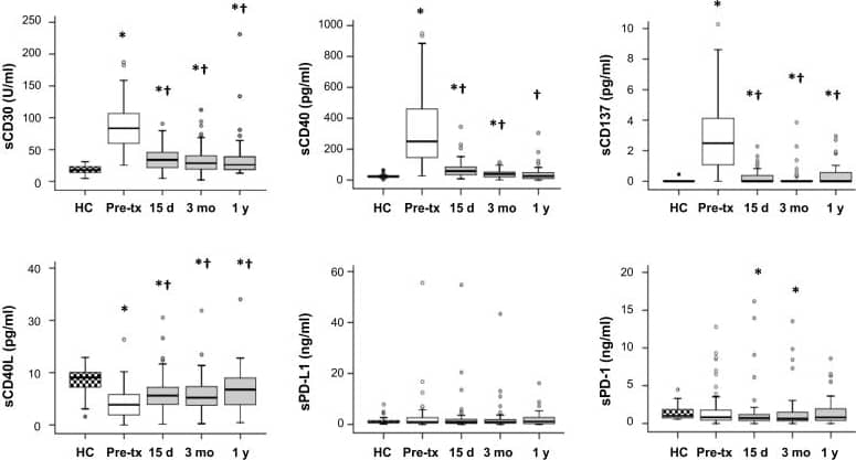
Applications: ELISA DevelopmentMelendreras S et al. (2014-12-05), Soluble co-signaling molecules predict long-term graft outcome in kidney-transplanted patients. PLoS ONE, 2014-19(12):e113396.
PMID: 25478957 -
Species: Primate - Macaca mulatta (Rhesus Macaque)
Sample Types: Whole Tissue
Applications: IHCHong J et al. (2014-06-06), Early lymphoid responses and germinal center formation correlate with lower viral load set points and better prognosis of simian immunodeficiency virus infection. J Immunol, 2014-0193(2):797-806.
PMID: 24907346 -
Species: Primate - Macaca mulatta (Rhesus Macaque)
Sample Types: Whole Tissue
Applications: IHC-P(2012-08-27), CD4 T follicular helper cell dynamics during SIV infection. J. Clin. Invest., 2012-0122(9):3281-94.
PMID: 22922258 -
Species: Human
Sample Types: Whole Tissue
Applications: IHCSchatton T et al. (2010-01-12), Modulation of T-cell activation by malignant melanoma initiating cells. Cancer Res., 2010-070(2):697-708.
PMID: 20068175 -
Species: Primate - Macaca mulatta (Rhesus Macaque),Primate - Cercocebus torquatus (Sooty Mangabey)
Sample Types: Whole Tissue
Applications: IHC-PEstes JD et al. (2008-05-15), Early resolution of acute immune activation and induction of PD-1 in SIV-infected sooty mangabeys distinguishes nonpathogenic from pathogenic infection in rhesus macaques. J. Immunol., 2008-0180(10):6798-807.
PMID: 18453600 -
Species: Primate - Macaca mulatta (Rhesus Macaque)
Sample Types: Whole Cells
Applications: NeutralizationOnlamoon N et al. (2008-02-05), Soluble PD-1 rescues the proliferative response of simian immunodeficiency virus-specific CD4 and CD8 T cells during chronic infection. Immunology, 2008-0124(2):277-93.
PMID: 18266718 -
Species: Human
Sample Types: Serum
Applications: ELISA DevelopmentWan B et al. (2006-12-15), Aberrant regulation of synovial T cell activation by soluble costimulatory molecules in rheumatoid arthritis. J. Immunol., 2006-1177(12):8844-50.
PMID: 17142787
There are no citations that match your criteria.
Customer Reviews for Human PD‑1 Antibody (6)
Customer Images
-
Application: Immunocytochemistry/ImmunofluorescenceSample Tested: Melanoma tissueSpecies: HumanVerified Customer | Posted 11/23/2020
-
Application: Western BlotSample Tested: 293T human embryonic kidney cell lineSpecies: HumanVerified Customer | Posted 07/23/2018293T cells were infected by lentivirus overexpression of control or PD-1 for 24 and 48 h. Total cell lysates were subjected to western blot. PVDF membrane were probed with 1 um/ml Human PD-1 Antibody (AF1086). A specific band was detected for PD-1 at approximately 43 kDa. This experiment was conducted under reducing conditions
-
Application: ELISASample Tested: Human recombinant antibodySpecies: HumanVerified Customer | Posted 03/07/2018
-
Application: ELISASample Tested: Human recombinant test antibodySpecies: HumanVerified Customer | Posted 09/29/2017
-
Application: Flow CytometrySample Tested: Peripheral blood mononuclear cells (PBMCs)Species: HumanVerified Customer | Posted 03/14/201710^6 Human PBMCs were probed with 0.5 ug of Goat anti-Human PD-1 antibody (red), or Goat Isotype Control IgG (black), followed by PE-conjugated anti-Goat secondary antibody.
-
Application: ImmunofluorescenceSample Tested: Mouse salivary gland (one injected with human T cells and one with no human T cells)Species: HumanVerified Customer | Posted 07/09/2016
There are no reviews that match your criteria.
Reconstitution Calculator
The reconstitution calculator allows you to quickly calculate the volume of a reagent to reconstitute your vial. Simply enter the mass of reagent and the target concentration and the calculator will determine the rest.