
Endothelial Cell Growth Media Best Seller
R&D Systems, part of Bio-Techne | Catalog # CCM027
Endothelial Cell Base Media plus Supplement


Key Product Details
Species
Human
Product Summary for Endothelial Cell Growth Media
Kit Summary
Media for the low-serum expansion of primary endothelial cells.
Key Benefits
- Unique and optimized formulation that promotes endothelial cell growth.
- Reduces cell senescence at higher passages
- Includes a single supplement featuring key growth factors and small molecules
- 250 mL bottle results in less wasted media
Why Use Pre-optimized, Low-serum Media to Grow Endothelial Cells?
Primary endothelial cells are commonly used for the study of developmental and disease mechanisms involving the blood vessels, such as angiogenesis. Endothelial cells are phenotypically characterized as CD31+, KDR+, and vWF+ cells.
R&D Systems® Endothelial Cell Growth Media is formulated to support the expansion of human primary endothelial cells under low-serum conditions, including HUVEC cell culture. Culturing endothelial cells under serum-free conditions allows for the researcher to have more controlled and defined system. This media has been tested for its ability to support CD31+, KDR+, and vWF+ human umbilical vein endothelial cells growth in vitro.
Kit Contents
This kit contains the following reagents to maintain and expand human and mouse endothelial cells in culture:
- Endothelial Cell Growth Base Media (250 mL)
- Endothelial Cell Growth Supplement (50X)
Containing optimized concentrations of:
- Human FGF basic
- Human LR3 IGF-1
- Human VEGF165
- Human EGF
- Heparin
- Hydrocortisone
- L-Ascorbic Acid
- Fetal Bovine Serum
Note: The components for this kit require different storage/shipping temperatures and will arrive in separate packaging.
Stability and Storage
Store in the dark at < -20 °C in a manual defrost freezer. Do not use past the expiration date.
Precaution
When handling biohazardous materials such as human cells, safe laboratory procedures should be followed and protective clothing should be worn.
Limitations
- FOR LABORATORY RESEARCH USE ONLY. NOT FOR USE IN DIAGNOSTIC PROCEDURES.
- The safety and efficacy of this product in diagnostic or other clinical uses has not been established.
- This reagent should not be used beyond the expiration date indicated on the label.
- Results may vary due to variations among cells derived from different donors.
Scientific Data Images for Endothelial Cell Growth Media

Endothelial Cell Growth Media Supports Cell Proliferation of Human Umbilical Cord Endothelial Cells (HUVECs).
One T25 flask of HUVECs (0.17 x 10⁶cells) were expanded over 10 passages using Complete Endothelial Cell Growth Media. Cells were passaged every 4-6 days. The histogram shows the cumulative number of HUVECs expanded over 10 passages if all cells were replated at each passage. This Endothelial Cell Growth Media functions as a HUVEC cell culture media.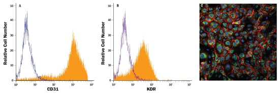
Human Umbilical Cord Endothelial Cells (HUVECs) Express Endothelial Cell Markers CD31, KDR, and vWF.
HUVECs were expanded in Complete Endothelial Cell Growth Media for 5 passages prior to staining for marker expression. (A,B) Flow cytometry analysis of endothelial cell marker expression. Filled histograms indicate cells stained with PE-conjugated Mouse Anti-CD31 (A; R&D Systems®, Catalog # FAB3567P) or PE-conjugated Mouse Anti-KDR (B; R&D Systems®, Catalog # FAB357P). Open histograms in A and B indicate staining with PE-conjugated Mouse IgG1 isotype control (R&D Systems®, Catalog # IC002P). (C) Immunocytochemistry analysis of endothelial cell marker expression. Cells were double stained with Mouse Anti-CD31 (red; R&D Systems®, Catalog # BBA7) and Rabbit Anti-Von Willebrand Factor (vWF) (green; Novus Biologicals®, Catalog # NB600-586). The nuclei were counterstained with DAPI (blue)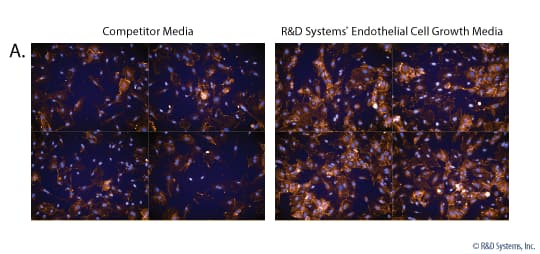
Increased HUVEC Survival and CD31 Expression Compared to Competitor Endothelial Cell Media.
HUVECs were expanded for 14 passages in either R&D Systems Endothelial Cell Growth Media (Catalog # CCM027) or competitor endothelial cell media. Cells were stained for CD31 expression using the Mouse Anti-Human CD31 Antibody (yellow; R&D Systems; Catalog # BBA7) and counterstained with DAPI (Catalog # 5748).A) Representative images of CD31 expression in p14 HUVECs grown in R&D Systems Media or competitor media. There is a greater density of cells and a higher expression of CD31 in HUVECs grown in R&D Systems Media.B) Histogram showing that at p14, there is a higher percentage of surviving cells that express CD31 in HUVECs grown in R&D Systems Media compared to competitor.

Formulation, Preparation, and Storage
Shipping
The product is shipped with polar packs. Upon receipt, store it immediately at the temperature recommended below.
Storage
Store the unopened product at 2 - 8 °C. Do not use past expiration date.
Product Documents for Endothelial Cell Growth Media
Product Specific Notices for Endothelial Cell Growth Media
For research use only
Loading...
Loading...
Loading...
Loading...