
Human Tie-2 PE-conjugated Antibody
R&D Systems, part of Bio-Techne | Catalog # FAB3131P

Scientific images may be resized, cropped, or adjusted for brightness or contrast for presentation purposes. These updates are limited to presentation and do not affect the underlying scientific interpretation of the data. Where available, additional source data may be provided upon request.
Conjugate
Catalog #
Key Product Details
Species Reactivity
Validated:
Human
Cited:
Human, Bovine, Primate - Macaca fascicularis (Crab-eating Monkey or Cynomolgus Macaque)
Applications
Validated:
Flow Cytometry
Cited:
Flow Cytometry
Label
Phycoerythrin (Excitation = 488 nm, Emission = 565-605 nm)
Antibody Source
Monoclonal Mouse IgG1 Clone # 83715
Product Specifications
Immunogen
Mouse myeloma cell line NS0-derived recombinant human Tie-2
Ala23-Lys745
Accession # AAA61139
Ala23-Lys745
Accession # AAA61139
Specificity
Detects human Tie-2 in direct ELISAs and Western blots. In direct ELISAs and Western blots, no cross-reactivity with recombinant human Tie‑1, recombinant mouse Tie-2, or recombinant zebrafish Tie-2 is observed.
Clonality
Monoclonal
Host
Mouse
Isotype
IgG1
Scientific Data Images for Human Tie-2 PE-conjugated Antibody
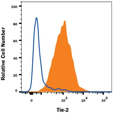
Detection of Tie‑2 in HUVEC Human Cells by Flow Cytometry.
HUVEC human umbilical vein endothelial cells were stained with Mouse Anti-Human Tie-2 PE-conjugated Monoclonal Antibody (Catalog # FAB3131P, filled histogram) or isotype control antibody (Catalog # IC002P, open histogram). View our protocol for Staining Membrane-associated Proteins.
Detection of Mouse Human Tie-2 PE-conjugated Antibody by Flow Cytometry
CSF1 up-regulates Tie2 receptor on CD14+ human monocytes.(A) CD14+ monocytes were isolated from whole blood using CD14+ microbeads. Cells were fixed and immunostained using anti-human Tie2 receptor antibody or isotype control antibody immediately following isolation (Freshly isolated) or after treated without (-CSF1) or with rhCSF1 (100 ng/ml) (+CSF1) for 24 hours. N = 10 per group and results represent the mean ± SEM of Tie2-positivity. (B) CD14+ monocytes treated with rhANG1 (100 ng/ml), rhANG2 (100 ng/ml) or a dose-response of rhCSF1 (0, 0.1, 1, 10, 100 ng/ml). ANG2 up-regulated Tie2 expression compared to ANG1 and CSF1 induces a dose-escalation of Tie2 on CD14+ monocytes. N = 10 per group and results represent the mean ± SEM of Tie2-positivity. (C) CD14+ monocytes were left untreated (Utx) or treated with rhANG2 (100 ng/ml) (ANG2), rhCSF1 (100 ng/ml) (CSF1), CSF1R neutralizing antibody alone, or pre-treated with the CSF1R Nab for 30 minutes prior to stimulation with rhCSF1 (100 ng/ml) (CSF1R NAb+CSF1) for 24 hours. ANG2- and CSF1-treatment significantly increased Tie2 expression while the CSF1R NAb abrogated this effect. N = 8 per group and results represent the mean ± SEM of Tie2-positivity by flow cytometry. (D) CD14+ monocytes were left untreated (Untreated), pre-treated with CSF1R NAb (40 µg or 80 µg) for 30 minutes then treated with rhCSF1 (100 ng/ml) (CSF1R NAb+CSF1), or with rhCSF1 (100 ng/ml) alone (CSF1) for 10 minutes. Western blot analysis indicates that the CSF1R NAb was effective at reducing Akt1 phosphorylation. Image collected and cropped by CiteAb from the following publication (https://pubmed.ncbi.nlm.nih.gov/24892425), licensed under a CC-BY license. Not internally tested by R&D Systems.
Detection of Mouse Human Tie-2 PE-conjugated Antibody by Flow Cytometry
CSF1 up-regulates Tie2 receptor on CD14+ human monocytes.(A) CD14+ monocytes were isolated from whole blood using CD14+ microbeads. Cells were fixed and immunostained using anti-human Tie2 receptor antibody or isotype control antibody immediately following isolation (Freshly isolated) or after treated without (-CSF1) or with rhCSF1 (100 ng/ml) (+CSF1) for 24 hours. N = 10 per group and results represent the mean ± SEM of Tie2-positivity. (B) CD14+ monocytes treated with rhANG1 (100 ng/ml), rhANG2 (100 ng/ml) or a dose-response of rhCSF1 (0, 0.1, 1, 10, 100 ng/ml). ANG2 up-regulated Tie2 expression compared to ANG1 and CSF1 induces a dose-escalation of Tie2 on CD14+ monocytes. N = 10 per group and results represent the mean ± SEM of Tie2-positivity. (C) CD14+ monocytes were left untreated (Utx) or treated with rhANG2 (100 ng/ml) (ANG2), rhCSF1 (100 ng/ml) (CSF1), CSF1R neutralizing antibody alone, or pre-treated with the CSF1R Nab for 30 minutes prior to stimulation with rhCSF1 (100 ng/ml) (CSF1R NAb+CSF1) for 24 hours. ANG2- and CSF1-treatment significantly increased Tie2 expression while the CSF1R NAb abrogated this effect. N = 8 per group and results represent the mean ± SEM of Tie2-positivity by flow cytometry. (D) CD14+ monocytes were left untreated (Untreated), pre-treated with CSF1R NAb (40 µg or 80 µg) for 30 minutes then treated with rhCSF1 (100 ng/ml) (CSF1R NAb+CSF1), or with rhCSF1 (100 ng/ml) alone (CSF1) for 10 minutes. Western blot analysis indicates that the CSF1R NAb was effective at reducing Akt1 phosphorylation. Image collected and cropped by CiteAb from the following publication (https://pubmed.ncbi.nlm.nih.gov/24892425), licensed under a CC-BY license. Not internally tested by R&D Systems.Applications for Human Tie-2 PE-conjugated Antibody
Application
Recommended Usage
Flow Cytometry
10 µL/106 cells
Sample: HUVEC human umbilical vein endothelial cells
Sample: HUVEC human umbilical vein endothelial cells
Reviewed Applications
Read 2 reviews rated 4 using FAB3131P in the following applications:
Spectra Viewer
Plan Your Experiments
Use our spectra viewer to interactively plan your experiments, assessing multiplexing options. View the excitation and emission spectra for our fluorescent dye range and other commonly used dyes.
Spectra Viewer
Formulation, Preparation, and Storage
Purification
Protein A or G purified from hybridoma culture supernatant
Formulation
Supplied in a saline solution containing BSA and Sodium Azide.
Shipping
The product is shipped with polar packs. Upon receipt, store it immediately at the temperature recommended below.
Stability & Storage
Protect from light. Do not freeze.
- 12 months from date of receipt, 2 to 8 °C as supplied.
Background: Tie-2
Tie-1/Tie (tyrosine kinase with Ig and EGF homology domains 1) and Tie-2/Tek comprise a receptor tyrosine kinase (RTK) subfamily with unique structural characteristics: two immunoglobulin-like domains flanking three epidermal growth factor (EGF)-like domains and followed by three fibronectin type III-like repeats in the extracellular region and a split tyrosine kinase domain in the cytoplasmic region. These receptors are expressed primarily on endothelial and hematopoietic progenitor cells and play critical roles in angiogenesis, vasculogenesis and hematopoiesis.
Human Tie-2 cDNA encodes a 1124 amino acid (aa) residue precursor protein with an 18 residue putative signal peptide, a 727 residue extracellular domain and a 354 residue cytoplasmic domain. Two ligands, angiopoietin-1 (Ang-1) and angiopoietin-2 (Ang-2), which bind Tie-2 with high-affinity have been identified. Ang-2 has been reported to act as an antagonist for Ang-1. Mice engineered to overexpress Ang-2 or to lack Ang-1 or Tie-2 display similar angiogenesis defects.
References
- Partanen, J. and D.J. Dumont (1999) Curr. Top. Microbiol. Immunol. 237:159.
- Takakura, N. et al. (1998) Immunity 9:677.
- Procopio, W. et al. (1999) J. Biol. Chem. 274:30196.
Long Name
Tyrosine Kinase with Immunoglobulin and Epidermal Growth Factor Homology Domains 2
Alternate Names
CD202b, TEK, Tie2
Entrez Gene IDs
Gene Symbol
TEK
UniProt
Additional Tie-2 Products
Product Documents for Human Tie-2 PE-conjugated Antibody
Product Specific Notices for Human Tie-2 PE-conjugated Antibody
For research use only
Loading...
Loading...
Loading...
Loading...
Loading...