
Human M-CSF PE-conjugated Antibody
R&D Systems, part of Bio-Techne | Catalog # IC2161P

Scientific images may be resized, cropped, or adjusted for brightness or contrast for presentation purposes. These updates are limited to presentation and do not affect the underlying scientific interpretation of the data. Where available, additional source data may be provided upon request.
Conjugate
Catalog #
Key Product Details
Validated by
Biological Validation
Species Reactivity
Validated:
Human
Cited:
Human
Applications
Validated:
Intracellular Staining by Flow Cytometry
Cited:
Flow Cytometry
Label
Phycoerythrin (Excitation = 488 nm, Emission = 565-605 nm)
Antibody Source
Monoclonal Mouse IgG2A Clone # 26786
Product Specifications
Immunogen
E. coli-derived recombinant human M-CSF
Specificity
Detects human M-CSF in flow cytometry.
Clonality
Monoclonal
Host
Mouse
Isotype
IgG2A
Scientific Data Images for Human M-CSF PE-conjugated Antibody
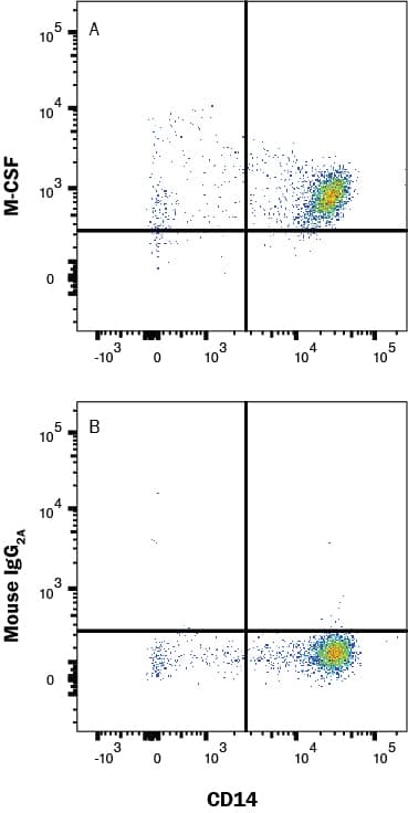
Detection of M-CSF in PBMC by Flow Cytometry
Human peripheral mononuclear cells (PBMC) were stained with Mouse Anti-Human CD14 Fluorescein-conjugated Monoclonal Antibody (Catalog # FAB3832F) and either (A) Mouse Anti-Human M-CSF PE-conjugated Monoclonal Antibody (Catalog # IC2161P) or (B) isotype control antibody (Catalog # IC003P). To facilitate intracellular staining, cells were fixed with Flow Cytometry Fixation Buffer (Catalog # FC004) and permeabilized with Flow Cytometry Permeabilization/Wash Buffer I (Catalog # FC005). View our protocol for Staining Intracellular Molecules.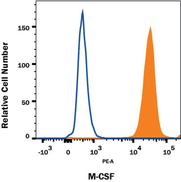
Detection of M‑CSF in Human Macrophages by Flow Cytometry.
Human macrophages treated with LPS and Recombinant Human TNF-a (210-TA) were stained with Mouse Anti-Human M-CSF PE-conjugated Monoclonal Antibody (Catalog # IC2161P, filled histogram) or isotype control antibody (IC003P, open histogram). To facilitate intracellular staining, cells were fixed with Flow Cytometry Fixation Buffer (FC004) and permeabilized with Flow Cytometry Permeabilization/Wash Buffer I (FC005). View our protocol for Staining Intracellular Molecules.Applications for Human M-CSF PE-conjugated Antibody
Application
Recommended Usage
Intracellular Staining by Flow Cytometry
10ul µL/106 cells
Sample: Human macrophages treated with LPS and Recombinant Human TNF-alpha (Catalog # 210-TA) and human PBMC's fixed with Flow Cytometry Fixation Buffer (Catalog # FC004) and permeabilized with Flow Cytometry Permeabilization/Wash Buffer I (Catalog # FC005)
Sample: Human macrophages treated with LPS and Recombinant Human TNF-alpha (Catalog # 210-TA) and human PBMC's fixed with Flow Cytometry Fixation Buffer (Catalog # FC004) and permeabilized with Flow Cytometry Permeabilization/Wash Buffer I (Catalog # FC005)
Spectra Viewer
Plan Your Experiments
Use our spectra viewer to interactively plan your experiments, assessing multiplexing options. View the excitation and emission spectra for our fluorescent dye range and other commonly used dyes.
Spectra Viewer
Formulation, Preparation, and Storage
Purification
Protein A or G purified from hybridoma culture supernatant
Formulation
Supplied in a saline solution containing BSA and Sodium Azide.
Shipping
The product is shipped with polar packs. Upon receipt, store it immediately at the temperature recommended below.
Stability & Storage
Store the unopened product at 2 - 8° C. Do not use past expiration date. Protect from light.
Background: M-CSF
Long Name
Macrophage Colony Stimulating Factor
Alternate Names
CSF-1, CSF1, Lanimostim, MCSF
Gene Symbol
CSF1
Additional M-CSF Products
Product Documents for Human M-CSF PE-conjugated Antibody
Product Specific Notices for Human M-CSF PE-conjugated Antibody
For research use only
Loading...
Loading...
Loading...
Loading...
Loading...