
Human CTLA-4 Antibody
R&D Systems, part of Bio-Techne | Catalog # AF-386-PB



Scientific images may be resized, cropped, or adjusted for brightness or contrast for presentation purposes. These updates are limited to presentation and do not affect the underlying scientific interpretation of the data. Where available, additional source data may be provided upon request.
Conjugate
Catalog #
Key Product Details
Validated by
Biological Validation
Species Reactivity
Validated:
Human
Cited:
Human, Rat, Bovine, Xenograft
Applications
Validated:
Western Blot, Flow Cytometry, Immunocytochemistry, CyTOF-ready
Cited:
Immunohistochemistry, Immunohistochemistry-Paraffin, Immunohistochemistry-Frozen, Flow Cytometry, In vivo assay
Label
Unconjugated
Antibody Source
Polyclonal Goat IgG
Product Specifications
Immunogen
S. frugiperda insect ovarian cell line Sf 21-derived recombinant human CTLA-4
Ala37-Phe162
Accession # Q6GR94
Ala37-Phe162
Accession # Q6GR94
Specificity
Detects human CTLA-4 in direct ELISAs and Western blots. In Western blots, approximately 25% cross-reactivity with recombinant mouse CTLA-4 is observed.
Clonality
Polyclonal
Host
Goat
Isotype
IgG
Scientific Data Images for Human CTLA-4 Antibody
Detection of Human CTLA‑4 by Western Blot.
Western blot shows lysates of NS0 mouse myeloma cell line either mock transfected or transfected with human CTLA-4. PVDF membrane was probed with 0.5 µg/mL of Goat Anti-Human CTLA-4 Antigen Affinity-purified Polyclonal Antibody (Catalog # AF-386-PB) followed by HRP-conjugated Anti-Goat IgG Secondary Antibody (Catalog # HAF017). A specific band was detected for CTLA-4 at approximately 50 kDa (as indicated). This experiment was conducted under reducing conditions and using Immunoblot Buffer Group 1.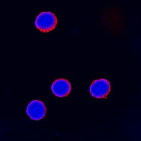
CTLA‑4 in Human Peripheral Blood Mononuclear Cells.
CTLA-4 was detected in immersion fixed human peripheral blood mononuclear cells treated with treated with PMA and calcium ionomycin using Goat Anti-Human CTLA-4 Antigen Affinity-purified Polyclonal Antibody (Catalog # AF-386-PB) at 15 µg/mL for 3 hours at room temperature. Cells were stained using the NorthernLights™ 557-conjugated Anti-Goat IgG Secondary Antibody (red; Catalog # NL001) and counterstained with DAPI (blue). Specific staining was localized to cell surfaces. View our protocol for Fluorescent ICC Staining of Non-adherent Cells.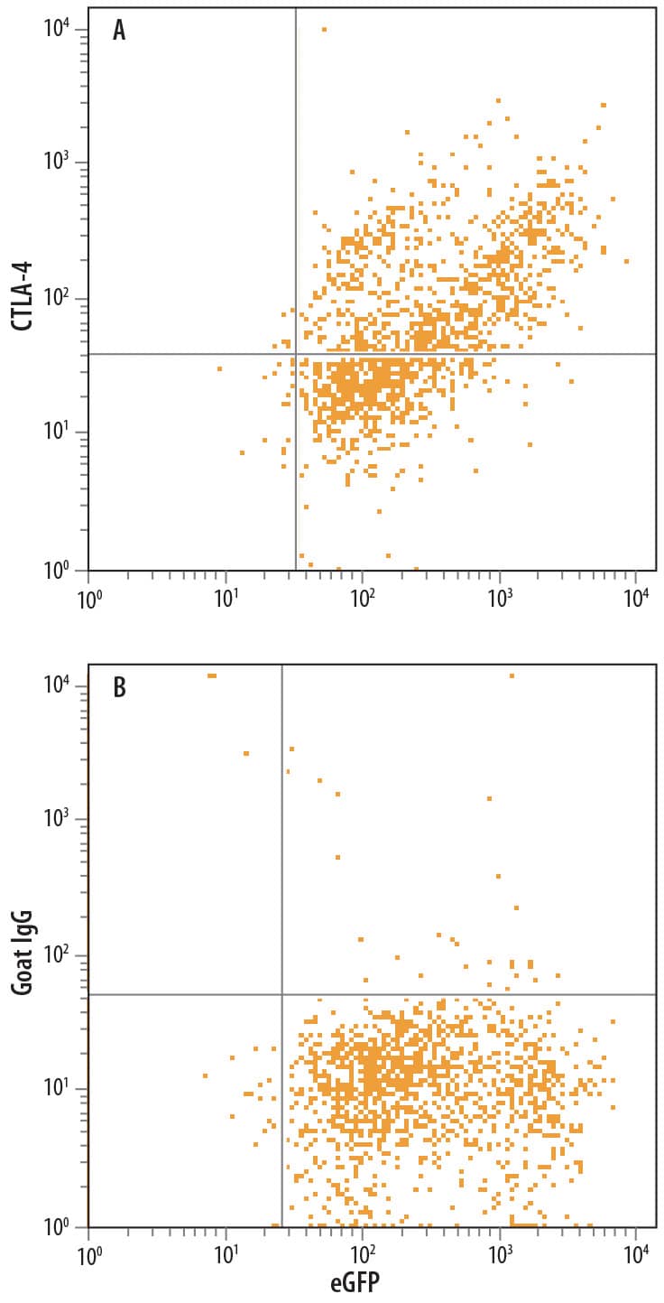
Detection of CTLA‑4 in NS0 Mouse Cell Line Co-transfected with CTLA-4 and eGFP by Flow Cytometry.
NS0 mouse myeloma cell line co-transfected with human CTLA-4 and eGFP was stained with either (A) Goat Anti-Human CTLA-4 Antigen Affinity-purified Polyclonal Antibody (Catalog # AF-386-PB) or (B) Normal Goat IgG Control (Catalog # AB-108-C) followed by Allophycocyanin-conjugated Anti-Goat IgG Secondary Antibody (Catalog # F0108).Applications for Human CTLA-4 Antibody
Application
Recommended Usage
CyTOF-ready
Ready to be labeled using established conjugation methods. No BSA or other carrier proteins that could interfere with conjugation.
Flow Cytometry
0.25 µg/106 cells
Sample: NS0 mouse myeloma cell line co-transfected with human CTLA-4 and eGFP
Sample: NS0 mouse myeloma cell line co-transfected with human CTLA-4 and eGFP
Immunocytochemistry
5-15 µg/mL
Sample: Immersion fixed human peripheral blood mononuclear cells treated with PMA and calcium ionomycin
Sample: Immersion fixed human peripheral blood mononuclear cells treated with PMA and calcium ionomycin
Western Blot
0.5 µg/mL
Sample: NS0 mouse myeloma cell line transfected with human CTLA-4
Sample: NS0 mouse myeloma cell line transfected with human CTLA-4
Reviewed Applications
Read 2 reviews rated 5 using AF-386-PB in the following applications:
Formulation, Preparation, and Storage
Purification
Antigen Affinity-purified
Reconstitution
Reconstitute at 0.2 mg/mL in sterile PBS. For liquid material, refer to CoA for concentration.
Formulation
Lyophilized from a 0.2 μm filtered solution in PBS with Trehalose. *Small pack size (SP) is supplied either lyophilized or as a 0.2 µm filtered solution in PBS.
Shipping
Lyophilized product is shipped at ambient temperature. Liquid small pack size (-SP) is shipped with polar packs. Upon receipt, store immediately at the temperature recommended below.
Stability & Storage
Use a manual defrost freezer and avoid repeated freeze-thaw cycles.
- 12 months from date of receipt, -20 to -70 °C as supplied.
- 1 month, 2 to 8 °C under sterile conditions after reconstitution.
- 6 months, -20 to -70 °C under sterile conditions after reconstitution.
Background: CTLA-4
CTLA-4 and CD28, together with their ligands B7-1 and B7-2, constitute one of the dominant costimulatory pathways that regulate T- and B-cell responses. CTLA-4 and CD28 are structurally homologous molecules that are members of the immunoglobulin (Ig) gene superfamily. Both CTLA-4 and CD28 are composed of a single
Ig V‑like extracellular domain, a transmembrane domain and an intracellular domain. CTLA-4 and CD28 are both expressed on the cell surface as disulfide-linked homodimers or as monomers. The genes encoding these two molecules are closely linked on human chromosome 2. CTLA-4 was originally identified as a gene that was specifically expressed by cytotoxic T lymphocytes. However, CTLA-4 transcripts have since been found in both Th1 and Th2, and CD4+ and CD8+ T cell clones. Whereas CD28 expression is constitutive on the surfaces of 95% of CD4+ T cells and 50% of CD8+ T cells and is down regulated upon T cell activation, CTLA-4 expression is upregulated rapidly following T cell activation and peaks approximately 24 hours following activation. Although both CTLA-4 and CD28 can bind to the same ligands, CTLA-4 binds to B7-1 and B7-2 with 20‑100‑fold higher affinity than CD28. The physiological role of CTLA-4 in T cell costimulation is currently being studied.
References
- Lenschow, D.J. et al. (1996) Annu. Rev. Immunol. 14:233.
- Hathcock, K.S. and R.J. Hodes (1996) Advances in Immunol. 62:131.
- Ward, S.G. (1996) Biochem. J. 318:361.
Long Name
Cytotoxic T-lymphocyte-associated Molecule 4
Alternate Names
CD152, CTLA4
Gene Symbol
CTLA4
UniProt
Additional CTLA-4 Products
Product Documents for Human CTLA-4 Antibody
Product Specific Notices for Human CTLA-4 Antibody
For research use only
Loading...
Loading...
Loading...
Loading...
Loading...
Loading...