
Human Cathepsin L Antibody
R&D Systems, part of Bio-Techne | Catalog # AF952

Scientific images may be resized, cropped, or adjusted for brightness or contrast for presentation purposes. These updates are limited to presentation and do not affect the underlying scientific interpretation of the data. Where available, additional source data may be provided upon request.
Conjugate
Catalog #
Key Product Details
Validated by
Knockout/Knockdown, Biological Validation
Species Reactivity
Validated:
Human
Cited:
Human, Mouse, Avian - Chicken, Xenograft
Applications
Validated:
Immunohistochemistry, Western Blot, ELISA Capture (Matched Antibody Pair), Immunoprecipitation
Cited:
Immunohistochemistry, Immunohistochemistry-Paraffin, Western Blot, Immunocytochemistry
Label
Unconjugated
Antibody Source
Polyclonal Goat IgG
Product Specifications
Immunogen
Mouse myeloma cell line NS0-derived recombinant human Cathepsin L
Glu113-Val333
Accession # P07711
Glu113-Val333
Accession # P07711
Specificity
Detects human Cathepsin L in ELISAs and Western blots.
Clonality
Polyclonal
Host
Goat
Isotype
IgG
Scientific Data Images for Human Cathepsin L Antibody
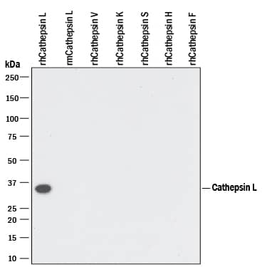
Detection of Recombinant Human Cathepsin L by Western Blot.
Western blot shows 10 ng of Recombinant Human Cathepsin L (952-CY), Recombinant Mouse Cathepsin L (1515-CY), Recombinant Human Cathepsin V (1080-CY), Recombinant Human Cathepsin K, Recombinant Human Cathepsin S (1183-CY), Recombinant Human Cathepsin H (7516-CY), and Recombinant Human Cathepsin F. PVDF Membrane was probed with 0.1 µg/mL of Goat Anti-Human Cathepsin L Antigen Affinity-purified Polyclonal Antibody (Catalog # AF952) followed by HRP-conjugated Anti-Goat IgG Secondary Antibody (HAF109). A specific band was detected for Cathepsin L at approximately 35 kDa (as indicated). This experiment was conducted under reducing conditions and using Immunoblot Buffer Group 3.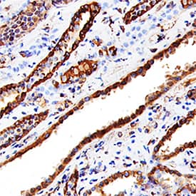
Cathepsin L in Human Kidney.
Cathepsin L was detected in immersion fixed paraffin-embedded sections of human kidney using Goat Anti-Human Cathepsin L Antigen Affinity-purified Polyclonal Antibody (Catalog # AF952) at 15 µg/mL overnight at 4 °C. Tissue was stained using the Anti-Goat HRP-DAB Cell & Tissue Staining Kit (brown; CTS008) and counterstained with hematoxylin (blue). Specific staining was localized to convoluted tubules. View our protocol for Chromogenic IHC Staining of Paraffin-embedded Tissue Sections.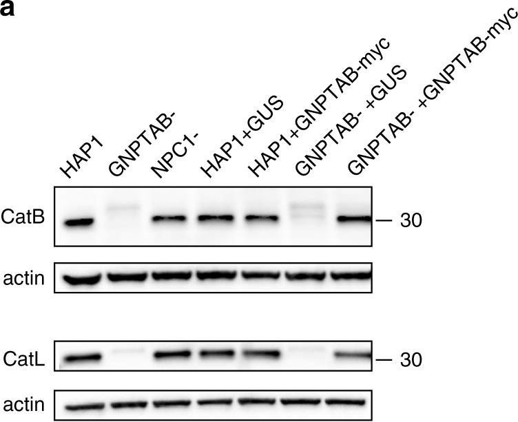
Detection of Human Cathepsin L by Western Blot
Cathepsins are reduced in GNPTAB- knockout cells, but restored upon GNPTAB reconstitution. a Immunoblotting of lysates from parental, knockout and reconstituted cells for CatB and CatL. The migration of a 30 kDa molecular mass marker is shown to the right. b CatB activity in lysates from parental HAP1, or knockout GNPTAB- or NPC1-cells. Cells were treated with cathepsin B inhibitor (Bi), cathepsin L inhibitor (Li), at 10 or 1 µM, or DMSO vehicle control for 1 h at 37 °C, before lysis and incubation with fluorescent CatB peptide substrate. After 1 h incubation at room temperature, fluorescence was measured. Data represent the mean ± s.d. of three technical replicates. A representative of three independent experiments is shown. c CatB activity in lysates from reconstituted cells. Lysates from transduced HAP1 or GNPTAB− cells were generated and tested for CatB activity, as for panel b. Data represent the mean ± s.d. of three technical replicates. A representative of two independent experiments is shown. For both panels b and c, statistical analysis was performed with a two-tailed Student’s t-test with significance shown as **** P ≤ 0.0001. Image collected and cropped by CiteAb from the following open publication (https://pubmed.ncbi.nlm.nih.gov/30655525), licensed under a CC-BY license. Not internally tested by R&D Systems.

Applications for Human Cathepsin L Antibody
Application
Recommended Usage
Immunohistochemistry
5-15 µg/mL
Sample: Immersion fixed paraffin-embedded sections of human kidney
Sample: Immersion fixed paraffin-embedded sections of human kidney
Immunoprecipitation
25 µg/mL
Sample: Conditioned cell culture medium spiked with Recombinant Human Cathepsin L (Catalog # 952-CY), see our available Western blot detection antibodies
Sample: Conditioned cell culture medium spiked with Recombinant Human Cathepsin L (Catalog # 952-CY), see our available Western blot detection antibodies
Western Blot
0.1 µg/mL
Sample: Recombinant Human Cathepsin L (Catalog # 952-CY)
Sample: Recombinant Human Cathepsin L (Catalog # 952-CY)
Human Cathepsin L Sandwich Immunoassay
Please Note: Optimal dilutions of this antibody should be experimentally determined.
Formulation, Preparation, and Storage
Purification
Antigen Affinity-purified
Reconstitution
Reconstitute at 0.2 mg/mL in sterile PBS. For liquid material, refer to CoA for concentration.
Formulation
Lyophilized from a 0.2 μm filtered solution in PBS with Trehalose. *Small pack size (SP) is supplied either lyophilized or as a 0.2 µm filtered solution in PBS.
Shipping
Lyophilized product is shipped at ambient temperature. Liquid small pack size (-SP) is shipped with polar packs. Upon receipt, store immediately at the temperature recommended below.
Stability & Storage
Use a manual defrost freezer and avoid repeated freeze-thaw cycles.
- 12 months from date of receipt, -20 to -70 °C as supplied.
- 1 month, 2 to 8 °C under sterile conditions after reconstitution.
- 6 months, -20 to -70 °C under sterile conditions after reconstitution.
Background: Cathepsin L
Cathepsin L is a lysosomal cysteine protease expressed in most eukaryotic cells. Cathepsin L is known to hydrolyze a number of proteins, including the proform of urokinase-type plasminogen activator, which is activated by Cathepsin L cleavage (1). Cathepsin L has also been shown to proteolytically inactivate alpha1-antitrypsin and secretory leucoprotease inhibitor, two major protease inhibitors of the respiratory tract (2). These observations, combined with the demonstration of increased Cathepsin L activity in the epithelial lining fluid of the lungs of emphysema patients, have led to the suggestion that the enzyme may be involved in the progression of this disease. Cathepsin L has also been identified as a major excreted protein of transformed fibroblasts, indicating the enzyme could be involved in malignant tumor growth (3). Human Cathepsin L activity is greatest under mildly acidic conditions, from pH 4.5‑6.5. The stability of the enzyme decreases at higher pH values.
References
- Goretzki, L. et al. (1992) FEBS Lett. 297:112.
- Taggart, C.C. et al. (2001) J. Biol. Chem. 276:33345.
- Gottesman, M.M. and F. Cabral (1981) Biochemistry 20:1659.
Alternate Names
CTSL, CTSL1
Gene Symbol
CTSL
UniProt
Additional Cathepsin L Products
Product Documents for Human Cathepsin L Antibody
Product Specific Notices for Human Cathepsin L Antibody
For research use only
Loading...
Loading...
Loading...
Loading...
Loading...