
Rat Lipocalin-2/NGAL Antibody
R&D Systems, part of Bio-Techne | Catalog # AF3508



Scientific images may be resized, cropped, or adjusted for brightness or contrast for presentation purposes. These updates are limited to presentation and do not affect the underlying scientific interpretation of the data. Where available, additional source data may be provided upon request.
Conjugate
Catalog #
Key Product Details
Species Reactivity
Validated:
Rat
Cited:
Human, Mouse, Rat, Transgenic Mouse
Applications
Validated:
Western Blot, ELISA Capture (Matched Antibody Pair), Immunoprecipitation
Cited:
Immunohistochemistry, Immunohistochemistry-Frozen, Western Blot, ELISA Detection, ELISA Development
Label
Unconjugated
Antibody Source
Polyclonal Goat IgG
Product Specifications
Immunogen
Mouse myeloma cell line NS0-derived recombinant rat Lipocalin-2/NGAL
Gln21-Asn198
Accession # P30152
Gln21-Asn198
Accession # P30152
Specificity
Detects rat Lipocalin-2/NGAL in ELISAs and detects rat and mouse Lipocalin-2/NGAL in Western blots. In sandwich ELISAs, less than 0.4% cross-reactivity with recombinant mouse (rm) Lipocalin-2/NGAL-2 is observed and less than 0.2% cross-reactivity with recombinant human (rh) Lipocalin-1/NGAL-1, rhLipocalin-2/NGAL-2 is observed.
Clonality
Polyclonal
Host
Goat
Isotype
IgG
Scientific Data Images for Rat Lipocalin-2/NGAL Antibody
Detection of Mouse and Rat Lipocalin-2/NGAL by Western Blot.
Western blot shows lysates of rat ovary tissue and mouse lung tissue. PVDF membrane was probed with 1 µg/mL of Mouse/Rat Lipocalin-2/NGAL Antigen Affinity-purified Polyclonal Antibody (Catalog # AF3508) followed by HRP-conjugated Anti-Goat IgG Secondary Antibody (Catalog # HAF019). A specific band was detected for Lipocalin-2/NGAL at approximately 25 kDa (as indicated). This experiment was conducted under reducing conditions and using Immunoblot Buffer Group 8.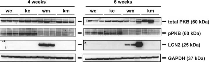
Detection of Mouse Lipocalin-2/NGAL by Western Blot
Visualization of expression of selected proteins by Western blot analysis. Protein extracts were prepared from livers of WT and Lcn2-/- mice that were subjected to indicated diets for 4 or 6 weeks (n=3 animals/group). Antibodies used are mentioned in the Section 2. GAPDH was used as the loading control. Image collected and cropped by CiteAb from the following publication (https://pubmed.ncbi.nlm.nih.gov/28725667), licensed under a CC-BY license. Not internally tested by R&D Systems.
Detection of Mouse Lipocalin-2/NGAL by Immunohistochemistry-Paraffin
Increased LCN2 expression in Esr1-deficient ovaries. Ovaries from wild-type (WT, n = 8) and Esr1 null mice (n = 6) were dissected and either embedded in paraffin or prepared for RNA analysis. (A) Hematoxylin and eosin (H & E) staining showed characteristic histological changes in Esr1-deficient compared to WT ovaries. (B) Immunohistochemical staining for LCN2 and ER alpha was performed. Sections were counterstained with hematoxylin (LCN2) or methyl green (ER alpha). Magnifications: 100×, 400×; scale bars: 100 µm, 50 µm. (C) Tissue samples were subjected to immunofluorescence double staining for LCN2 and STAR. Magnifications: 100×; scale bars: 100 µm. (D) Lcn2 and Star expression was analyzed in ovarian tissues using RT-qPCR. Data are displayed as means ± SD. In case of normal distribution, Student’s t-tests were chosen for statistical analysis, otherwise a Mann–Whitney test was performed. Significant differences between groups are marked with asterisks: *** p < 0.001. Image collected and cropped by CiteAb from the following open publication (https://pubmed.ncbi.nlm.nih.gov/37298232), licensed under a CC-BY license. Not internally tested by R&D Systems.


Applications for Rat Lipocalin-2/NGAL Antibody
Application
Recommended Usage
Immunoprecipitation
25 µg/mL
Sample: Conditioned cell culture medium spiked with Recombinant Rat Lipocalin-2/NGAL (Catalog # 3508-LC), see our available Western blot detection antibodies
Sample: Conditioned cell culture medium spiked with Recombinant Rat Lipocalin-2/NGAL (Catalog # 3508-LC), see our available Western blot detection antibodies
Western Blot
1 µg/mL
Sample: Rat ovary tissue and mouse lung tissue
Sample: Rat ovary tissue and mouse lung tissue
Rat Lipocalin-2 Sandwich Immunoassay
Please Note: Optimal dilutions of this antibody should be experimentally determined.
Reviewed Applications
Read 1 review rated 5 using AF3508 in the following applications:
Formulation, Preparation, and Storage
Purification
Antigen Affinity-purified
Reconstitution
Reconstitute at 0.2 mg/mL in sterile PBS. For liquid material, refer to CoA for concentration.
Formulation
Lyophilized from a 0.2 μm filtered solution in PBS with Trehalose. *Small pack size (SP) is supplied either lyophilized or as a 0.2 µm filtered solution in PBS.
Shipping
Lyophilized product is shipped at ambient temperature. Liquid small pack size (-SP) is shipped with polar packs. Upon receipt, store immediately at the temperature recommended below.
Stability & Storage
Use a manual defrost freezer and avoid repeated freeze-thaw cycles.
- 12 months from date of receipt, -20 to -70 °C as supplied.
- 1 month, 2 to 8 °C under sterile conditions after reconstitution.
- 6 months, -20 to -70 °C under sterile conditions after reconstitution.
Background: Lipocalin-2/NGAL
Lipocalin-2, also known as neutrophil gelatinase-associated Lipocalin and uterocalin (NGAL), has been implicated in a variety of processes including cell differentiation, tumorigenesis, and apoptosis (1‑3). It binds a bacterial catecholate sidropore bound to ferric ion such as enterobactin with a subnanomolar dissociation constant (KD = 0.41 nM) (4). The bound ferric enterobactin complex breaks down slowly in a month into dihydroxybenzoyl serine and dihydroxybenzoic acid (DHBA). It also binds to a ferric DHBA complex with much less KD values (7.9 nM) (4). Secretion of Lipocalin-2 in immune cells increases by stimulation of Toll-like receptor as a acute phase response to infection. As a result, it acts as a potent bacteriostatic reagents by sequestering iron (5). Moreover, Lipocalin-2 can alter the invasive and metastatic behavior of Ras-transformed breast cancer cells in vitro and in vivo by reversing epithelial to mesenchymal transition inducing activity of Ras, through restoration of E-cadherin expression, via effects on the Ras-MAPK signaling pathway (6). In the kidney, Lipocalin-2-mediated iron trafficking may be involved in protection from renal injury, and it has been implicated as a marker for early kidney failure (7, 8).
References
- Kjeldsen L, et al. (2000) Biochim Biophys Acta. 1482:272.
- Devireddy, L.R. et al. (2001) Science 293:829.
- Yang, M.B. et al. (2002) Mol. Cell. 10:1045.
- Goetz, D.H. et al. (2002) Mol. Cell 10:1033.
- Flo, T.H. et al. (2004) Nature 432:917.
- Hanai, J. et al. (2005) J. Biol. Chem. 280:13641.
- Mori, K. et al. (2005) J. Clin. Invest. 115:610.
- Mishra, J. et al. (2005) Lancet 365:1231.
Long Name
Neutrophil Gelatinase-associated Lipocalin
Alternate Names
24p3, LCN2, Lipocalin2, MSFI, NGAL, Oncogene 24p3, p25, Siderocalin, Uterocalin
Gene Symbol
LCN2
UniProt
Additional Lipocalin-2/NGAL Products
Product Documents for Rat Lipocalin-2/NGAL Antibody
Product Specific Notices for Rat Lipocalin-2/NGAL Antibody
For research use only
Loading...
Loading...
Loading...
Loading...
Loading...