Mouse PD-L1/B7-H1 Antibody
R&D Systems, part of Bio-Techne | Catalog # MAB11630


Key Product Details
Species Reactivity
Applications
Label
Antibody Source
Product Specifications
Immunogen
Phe19-His239
Accession # Q9EP73
Specificity
Clonality
Host
Isotype
Scientific Data Images for Mouse PD-L1/B7-H1 Antibody
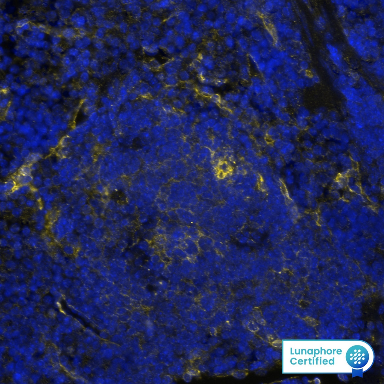
Detection of PD-L1/B7-H1 in Mouse Spleen via Multiplex Immunofluorescence staining on COMET™
PD-L1/B7-H1 was detected in immersion fixed paraffin-embedded sections of mouse spleen using Rabbit Anti-Mouse PD-L1/B7-H1 Recombinant Monoclonal Antibody (Catalog # MAB11630) at 5ug/mL at 37 ° Celsius for 4 minutes. Before incubation with the primary antibody, tissue underwent an all-in-one dewaxing and antigen retrieval preprocessing using PreTreatment Module (PT Module) and Dewax and HIER Buffer H (pH 9). Tissue was stained using the Alexa Fluor™ Plus 647 Goat anti-Rabbit IgG Secondary Antibody at 1:200 at 37 ° Celsius for 2 minutes. (Yellow; Lunaphore Catalog # DR647RB) and counterstained with DAPI (blue; Lunaphore Catalog # DR100). Specific staining was localized to the membrane. Protocol available in COMET™ Panel Builder.Detection of PD-L1/B7-H1 in Mouse Thymus.
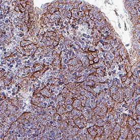
PD-L1/B7-H1 was detected in immersion fixed paraffin-embedded sections of mouse thymus using Rabbit Anti-Mouse PD-L1/B7-H1 Monoclonal Antibody (Catalog # MAB11630) at 0.075 µg/ml for 1 hour at room temperature followed by incubation with the Anti-Rabbit IgG VisUCyte™ HRP Polymer Antibody (Catalog # VC003) or the HRP-conjugated Anti-Rabbit IgG Secondary Antibody (Catalog # HAF008). Before incubation with the primary antibody, tissue was subjected to heat-induced epitope retrieval using VisUCyte Antigen Retrieval Reagent-Basic (Catalog # VCTS021). Tissue was stained using DAB (brown) and counterstained with hematoxylin (blue). Specific staining was localized to the membrane. View our protocol for IHC Staining with VisUCyte HRP Polymer Detection Reagents.Detection of PD-L1/B7-H1 in Mouse Thymus.
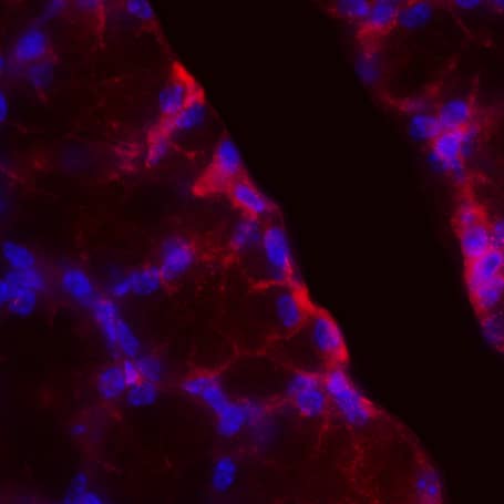
PD-L1/B7-H1 was detected in perfusion fixed frozen sections of mouse thymus using Rabbit Anti-Mouse PD-L1/B7-H1 Monoclonal Antibody (Catalog # MAB11630) at 1 µg/ml overnight at 4 °C. Tissue was stained using the NorthernLights™ 557-conjugated Anti-Rabbit IgG Secondary Antibody (red; Catalog # NL004) and counterstained with hematoxylin (blue). Specific staining was localized to the membrane. View our protocol for Fluorescent IHC Staining of Frozen Tissue Sections.Applications for Mouse PD-L1/B7-H1 Antibody
Immunohistochemistry
Sample: Perfusion fixed frozen sections of mouse thymus and immersion fixed paraffin-embedded sections of mouse thymus
Multiplex Immunofluorescence
Sample: Immersion fixed paraffin-embedded sections of Mouse Spleen
Formulation, Preparation, and Storage
Purification
Reconstitution
Formulation
Shipping
Stability & Storage
- 12 months from date of receipt, -20 to -70 °C as supplied.
- 1 month, 2 to 8 °C under sterile conditions after reconstitution.
- 6 months, -20 to -70 °C under sterile conditions after reconstitution.
Background: PD-L1/B7-H1
Mouse B7 homolog 1(B7-H1), also called programmed death ligand 1 (PD-L1) and programmed cell death 1 ligand 1 (PDCD1L1), is a member of the B7 family of proteins that provide signals for regulating T-cell activation and tolerance (1‑4). Other family members include B7-1, B7-2, B7-H2, B7-H3 and PD-L2. B7 proteins are immunoglobulin (Ig) superfamily members with extracellular Ig-V-like and Ig-C-like domains and a short cytoplasmic region. Among the family members, they share from 20‑40% amino acid (aa) sequence identity. The cloned mouse B7-H1/PD-L1 cDNA encodes a 290 aa type I membrane precursor protein with a putative 18 aa signal peptide, a 220 aa extracellular region containing one V-like and one C-like Ig domain, a 22 aa transmembrane region, and a 30 aa cytoplasmic domain. Mouse and human B7-H1/PD-L1 share approximately 70% aa sequence identity. B7-H1/PD-L1 is one of two ligands for programmed death-1 (PD-1), a member of the CD28 family of immunoreceptors. The other identified ligand is PD-L2. Mouse B7-H1/PD-L1 and PD-L2 share approximately 34% aa sequence identity and have similar functions. B7-H1/PD-L1 is constitutively expressed in various lymphoid and non-lymphoid organs including placenta, heart, pancreas, lung, liver, and endothelium
(1‑4). The expression of B7-H1/PD-L1 is detected on B cells, T cells, monocytes, dendritic cells and thymic epithelial cells. IFN-gamma treatment induces B7-H1/PD-L1 expression in monocytes, dendritic cells, and endothelial cells. B7-H1/PD-L1 expression is also upregulated in a variety of tumor cell lines. On previously activated T cells, B7-H1/PD-L1 interaction with PD-1 inhibits TCR-mediated proliferation and cytokine production, suggesting an inhibitory role in regulating immune responses. In contrast, a costimulatory function for the PD-1 ligands on resting T cells has also been reported (1‑4).
References
- Tamura, H. et al. (2001) Blood 97:1809.
- Freeman, G. et al. (2000) J. Exp. Med. 192:1027.
- Sharpe, A.H. and G. J. Freeman (2002) Nat. Rev. Immunol. 2:116.
- Coyle, A. and J. Gutierrez-Ramos (2001) Nat. Immunol. 2:203.
Long Name
Alternate Names
Entrez Gene IDs
Gene Symbol
UniProt
Additional PD-L1/B7-H1 Products
Product Documents for Mouse PD-L1/B7-H1 Antibody
Product Specific Notices for Mouse PD-L1/B7-H1 Antibody
For research use only

