
Human Phospho-PDGF R alpha (Y762) Antibody
R&D Systems, part of Bio-Techne | Catalog # AF21141


Scientific images may be resized, cropped, or adjusted for brightness or contrast for presentation purposes. These updates are limited to presentation and do not affect the underlying scientific interpretation of the data. Where available, additional source data may be provided upon request.
Key Product Details
Validated by
Biological Validation
Species Reactivity
Validated:
Human
Cited:
Human
Applications
Validated:
Immunohistochemistry, Western Blot
Cited:
Western Blot
Label
Unconjugated
Antibody Source
Polyclonal Rabbit IgG
Product Specifications
Immunogen
Phosphopeptide containing human PDGF R alpha Y762 site
Specificity
Detects human PDGF R alpha when phosphorylated at Y762.
Clonality
Polyclonal
Host
Rabbit
Isotype
IgG
Scientific Data Images for Human Phospho-PDGF R alpha (Y762) Antibody
Detection of Human Phospho-PDGF R alpha (Y762) by Western Blot.
Western blot shows lysates of human foreskin fibroblast untreated (-) or treated (+) with 100 ng/mL Recombinant Human PDGF-AA (Catalog # 221-AA) or Recombinant Human PDGF-BB (Catalog # 220-BB) for 10 minutes. PVDF membrane was probed with 0.5 µg/mL of Human Phospho-PDGF Ra (Y762) Antigen Affinity-purified Polyclonal Antibody (Catalog # AF21141), followed by HRP-conjugated Anti-Rabbit IgG Secondary Antibody (Catalog # HAF008). A specific band was detected for Phospho-PDGF Ra (Y762) at approximately 180 kDa (as indicated). This experiment was conducted under reducing conditions and using Immunoblot Buffer Group 1.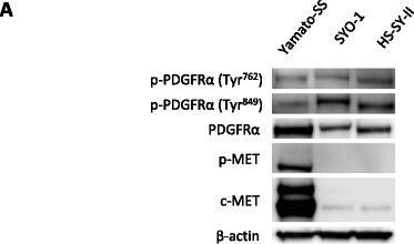
Detection of Phospho-PDGF R alpha (Y762) by Western Blot
c-MET and PDGFR alpha signals are crucial for the proliferation of SS cells. a Phosphorylation status of RTKs in 3 SS cell lines. b Yamato-SS (3 × 10³), SYO-1 (5 × 10³) and HS-SY-II (1 × 10⁴) cells were transfected with siRNAs against c-MET. Transfected cells were cultured for 96 h and relative cell proliferation rates were measured using a WST-1 assay. Bars represent the SD. * p < 0.05. c Growth of Yamato-SS (3 × 10³), SYO-1 (5 × 10³) and HS-SY-II (1 × 10⁴) cells transfected with siRNAs against PDGFR alpha. Relative cell proliferation rates were determined using a WST-1 assay after 96 h. Bars represent the SD. * p < 0.05 Image collected and cropped by CiteAb from the following open publication (https://pubmed.ncbi.nlm.nih.gov/28511645), licensed under a CC-BY license. Not internally tested by R&D Systems.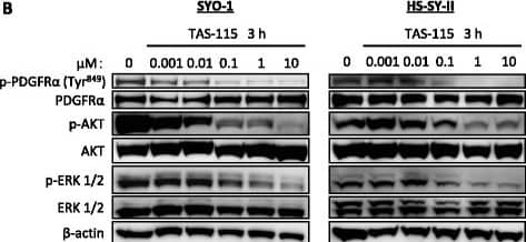
Detection of Phospho-PDGF R alpha (Y762) by Western Blot
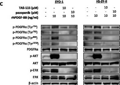
TAS-115 suppresses phosphorylation of c-MET and PDGFR alpha, as well as their downstream effectors. a Yamato-SS cells (c-MET-dependent SS cells) were treated with 0.001–10 μM of TAS-115 or control (0.1% DMSO) for 3 h. b SYO-1 and HS-SY-II cells (PDGFR alpha-dependent SS cells) were treated with 0.001–10 μM of TAS-115 or control (0.1% DMSO) for 3 h Image collected and cropped by CiteAb from the following open publication (https://pubmed.ncbi.nlm.nih.gov/28511645), licensed under a CC-BY license. Not internally tested by R&D Systems.Applications for Human Phospho-PDGF R alpha (Y762) Antibody
Application
Recommended Usage
Immunohistochemistry
5-15 µg/mL
Sample: Immersion fixed paraffin-embedded sections of human breast cancer tissue
Sample: Immersion fixed paraffin-embedded sections of human breast cancer tissue
Western Blot
0.5 µg/mL
Sample: Human foreskin fibroblast treated with Recombinant Human PDGF-BB (Catalog # 220-BB)
Sample: Human foreskin fibroblast treated with Recombinant Human PDGF-BB (Catalog # 220-BB)
Reviewed Applications
Read 1 review rated 5 using AF21141 in the following applications:
Formulation, Preparation, and Storage
Purification
Antigen Affinity-purified
Reconstitution
Reconstitute at 0.2 mg/mL in sterile PBS. For liquid material, refer to CoA for concentration.
Formulation
Lyophilized from a 0.2 μm filtered solution in PBS with Trehalose. *Small pack size (SP) is supplied either lyophilized or as a 0.2 µm filtered solution in PBS.
Shipping
Lyophilized product is shipped at ambient temperature. Liquid small pack size (-SP) is shipped with polar packs. Upon receipt, store immediately at the temperature recommended below.
Stability & Storage
Use a manual defrost freezer and avoid repeated freeze-thaw cycles.
- 12 months from date of receipt, -20 to -70 °C as supplied.
- 1 month, 2 to 8 °C under sterile conditions after reconstitution.
- 6 months, -20 to -70 °C under sterile conditions after reconstitution.
Background: PDGF R alpha
PDGF is a major serum mitogen that can exist as a homo- or heterodimeric protein consisting of disulfide-linked PDGF-A and PDGF-B chains. The PDGF-AA, PDGF‑BB and PDGF-AB isoforms have been shown to bind to two distinct cell surface PDGF receptors with different affinities. Whereas PDGF R alpha binds all three PDGF isoforms with high affinity, PDGF R beta binds PDGF-BB and AB, but not PDGF-AA. Both PDGF R alpha and PDGF R beta are members of the class III subfamily of receptor tyrosine kinases (RTK) that also includes the receptors for M-CSF, SCF and Flt3 ligand. All class III RTKs are characterized by the presence of five immunoglobulin-like domains in their extracellular region and a split kinase domain in their intracellular region. PDGF binding induces receptor homo- and heterodimerization and signal transduction. The expression of the alpha and beta receptors is independently regulated in various cell types. Only PDGF R alpha is expressed in oligodendrocyte progenitor cells, mesothelial cell and liver endothelial cells. Soluble PDGF-R alpha has been detected in cell conditioned medium and human plasma. Recombinant soluble PDGF R alpha binds PDGF with high affinity and is a potent PDGF antagonist (1).
References
- Heldin, C.H. and L. Claesson-Welsh (1994) Guidebook to Cytokines and Their Receptors, Nicola, N.A. (ed) Oxford University Press, New York, NY p. 202.
Long Name
Platelet-derived Growth Factor Receptor alpha
Alternate Names
CD140a, PDGFR alpha, PDGFRA
Gene Symbol
PDGFRA
Additional PDGF R alpha Products
Product Documents for Human Phospho-PDGF R alpha (Y762) Antibody
Product Specific Notices for Human Phospho-PDGF R alpha (Y762) Antibody
For research use only
Loading...
Loading...
Loading...
Loading...
Loading...