
Human ICAM-1/CD54 Antibody
R&D Systems, part of Bio-Techne | Catalog # AF720



Conjugate
Catalog #
Key Product Details
Validated by
Knockout/Knockdown, Biological Validation
Species Reactivity
Validated:
Human
Cited:
Human
Applications
Validated:
Knockout Validated, Western Blot, Adhesion Blockade, Immunocytochemistry
Cited:
Western Blot, Neutralization, Immunocytochemistry, Bioassay
Label
Unconjugated
Antibody Source
Polyclonal Sheep IgG
Product Specifications
Immunogen
Chinese hamster ovary cell line CHO-derived recombinant human ICAM-1
Extracellular domain
Extracellular domain
Specificity
Selected for its ability to block the adhesion of HSB‑2 cells to immobilized recombinant human (rh) ICAM‑1. In direct ELISAs, approximately 5% cross-reactivity with rhICAM-3 is observed and less than 2% cross-reactivity with rhICAM-2, rhICAM-4, rhICAM-5, recombinant rat ICAM‑1, recombinant mouse ICAM‑1, and rhVCAM-1 is observed.
Clonality
Polyclonal
Host
Sheep
Isotype
IgG
Endotoxin Level
<0.20 EU per 1 μg of the antibody by the LAL method.
Scientific Data Images for Human ICAM-1/CD54 Antibody
Detection of Human ICAM‑1/CD54 by Western Blot.
Western blot shows lysates of HUVEC human umbilical vein endothelial cells and Ramos human Burkitt's lymphoma cell line untreated (-) or treated (+) with 10 ng/mL Recombinant Human TNF-a (Catalog # 210-TA) for 24 hours. PVDF membrane was probed with 0.2 µg/mL of Sheep Anti-Human ICAM-1/CD54 Antigen Affinity-purified Polyclonal Antibody (Catalog # AF720) followed by HRP-conjugated Anti-Sheep IgG Secondary Antibody (Catalog # HAF016). A specific band was detected for ICAM-1/CD54 at approximately 90 kDa (as indicated). This experiment was conducted under reducing conditions and using Immunoblot Buffer Group 1.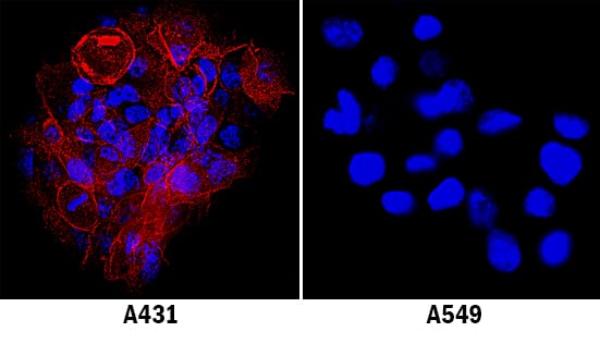
ICAM‑1/CD54 in A431 Human Cell Line.
ICAM-1/CD54 was detected in immersion fixed A431 human epithelial carcinoma cell line (left panel; positive stain) and A549 human lung carcinoma cell line (right panel; negative stain) using Sheep Anti-Human ICAM-1/CD54 Antigen Affinity-purified Polyclonal Antibody (Catalog # AF720) at 1.7 µg/mL for 3 hours at room temperature. Cells were stained using the NorthernLights™ 557-conjugated Anti-Sheep IgG Secondary Antibody (red; Catalog # NL010) and counterstained with DAPI (blue). Specific staining was localized to plasma membrane. View our protocol for Fluorescent ICC Staining of Cells on Coverslips.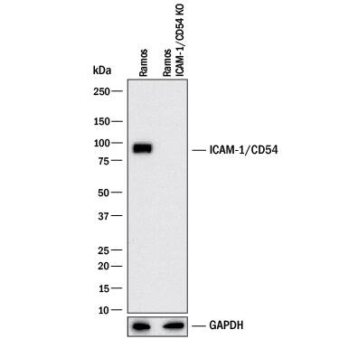
Western Blot Shows Human ICAM-1/CD54 Specificity by Using Knockout Cell Line.
Western blot shows lysates of Ramos human Burkitt's lymphoma cell line and human ICAM-1/CD54 Ramos human Burkitt's lymphoma cell line (KO). PVDF membrane was probed with 0.2 µg/mL of Sheep Anti-Human ICAM-1/CD54 Antigen Affinity-purified Polyclonal Antibody (Catalog # AF720) followed by HRP-conjugated Anti-Sheep IgG Secondary Antibody (HAF016). A specific band was detected for ICAM-1/CD54 at approximately 90 kDa (as indicated) in the parental Ramos human Burkitt's lymphoma cell line, but is not detectable in knockout Ramos human Burkitt's lymphoma cell line. GAPDH (Catalog # AF5718) is shown as a loading control. This experiment was conducted under reducing conditions and using Western Blot Buffer Group 1.Applications for Human ICAM-1/CD54 Antibody
Application
Recommended Usage
Adhesion Blockade
The adhesion of HSB2 human peripheral blood acute lymphoblastic leukemia cells (5 x 10⁴ cells/well) to immobilized recombinant human ICAM-1/Fc Chimera (12.5 μg/mL, 100 µL/well) was maximally inhibited (80‑100%) by 25 μg/mL of the antibody.
Immunocytochemistry
1-15 µg/mL
Sample: Immersion fixed A431 human epithelial carcinoma cell line
Sample: Immersion fixed A431 human epithelial carcinoma cell line
Knockout Validated
ICAM‑1/CD54 is specifically detected in the parental Ramos human Burkitt's lymphoma cell line but is not detectable in knockout Ramos human Burkitt's lymphoma cell line.
Western Blot
0.2 µg/mL
Sample: HUVEC human umbilical vein endothelial cells treated with Recombinant Human TNF‑ alpha (Catalog # 210-TA) and Ramos human Burkitt's lymphoma cell line
Sample: HUVEC human umbilical vein endothelial cells treated with Recombinant Human TNF‑ alpha (Catalog # 210-TA) and Ramos human Burkitt's lymphoma cell line
Reviewed Applications
Read 2 reviews rated 4.5 using AF720 in the following applications:
Formulation, Preparation, and Storage
Purification
Antigen Affinity-purified
Reconstitution
Reconstitute at 0.2 mg/mL in sterile PBS. For liquid material, refer to CoA for concentration.
Formulation
Lyophilized from a 0.2 μm filtered solution in PBS with Trehalose. *Small pack size (SP) is supplied either lyophilized or as a 0.2 µm filtered solution in PBS.
Shipping
Lyophilized product is shipped at ambient temperature. Liquid small pack size (-SP) is shipped with polar packs. Upon receipt, store immediately at the temperature recommended below.
Stability & Storage
Use a manual defrost freezer and avoid repeated freeze-thaw cycles.
- 12 months from date of receipt, -20 to -70 °C as supplied.
- 1 month, 2 to 8 °C under sterile conditions after reconstitution.
- 6 months, -20 to -70 °C under sterile conditions after reconstitution.
Background: ICAM-1/CD54
Intercellular Adhesion Molecule-1 (ICAM-1) binds the leukocyte integrins LFA-1 and Mac-1. ICAM-1 expression is weak on leukocytes, epithelial and resting endothelial cells, as well as some other cell types, but expression can be stimulated by IFN-gamma, TNF-alpha, IL-1 beta and LPS. Soluble ICAM-1 is found in a biologically active form in serum, probably as a result of proteolytic cleavage from the cell surface, and is elevated in patients with various inflammatory syndromes such as septic shock, LAD, cancer and transplantation.
Long Name
Intercellular Adhesion Molecule 1
Alternate Names
CD54, ICAM1
Gene Symbol
ICAM1
Additional ICAM-1/CD54 Products
Product Documents for Human ICAM-1/CD54 Antibody
Product Specific Notices for Human ICAM-1/CD54 Antibody
For research use only
Loading...
Loading...
Loading...
Loading...
Loading...