
Human Dectin-1/CLEC7A Antibody
R&D Systems, part of Bio-Techne | Catalog # MAB1859
Clone 259931 was used by HLDA to establish CD designation


Scientific images may be resized, cropped, or adjusted for brightness or contrast for presentation purposes. These updates are limited to presentation and do not affect the underlying scientific interpretation of the data. Where available, additional source data may be provided upon request.
Conjugate
Catalog #
Key Product Details
Species Reactivity
Validated:
Human
Cited:
Human, Mouse
Applications
Validated:
Blockade of Receptor-ligand Interaction, Flow Cytometry, Immunocytochemistry, CyTOF-ready
Cited:
Immunohistochemistry, Immunohistochemistry-Paraffin, Western Blot, Neutralization, Flow Cytometry, Immunocytochemistry, Immunoprecipitation, Bioassay, Blocking, Functional Assay
Label
Unconjugated
Antibody Source
Monoclonal Mouse IgG2B Clone # 259931
Product Specifications
Immunogen
Mouse myeloma cell line NS0-derived recombinant human Dectin‑1/CLEC7A
Thr66-Met201
Accession # NP_072092
Thr66-Met201
Accession # NP_072092
Specificity
Detects human Dectin‑1/CLEC7A in direct ELISAs. In directs ELISAs, less than 10% cross-reactivity with recombinant human (rh) DLEC and no cross-reactivity with recombinant mouse Dectin-1 or rhDectin-2 is observed.
Clonality
Monoclonal
Host
Mouse
Isotype
IgG2B
Endotoxin Level
<0.10 EU per 1 μg of the antibody by the LAL method.
Scientific Data Images for Human Dectin-1/CLEC7A Antibody
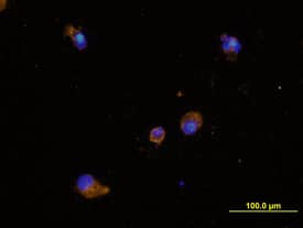
Dectin‑1/CLEC7A in Human Dendritic Cells.
Dectin-1/CLEC7A was detected in immersion fixed mature human dendritic cells using Mouse Anti-Human Dectin-1/CLEC7A Monoclonal Antibody (Catalog # MAB1859) at 10 µg/mL for 3 hours at room temperature. Cells were stained using the NorthernLights™ 557-conjugated Anti-Mouse IgG Secondary Antibody (yellow; Catalog # NL007) and counterstained with DAPI (blue). View our protocol for Fluorescent ICC Staining of Non-adherent Cells.
Detection of hcxtnxqqrfkwbdd2uwhc.jpg by Flow Cytometry
Glycosylation of the stalk region of hDectin-1 mediates recognition by CLEC-2.(A) Schematic representations of mouse Dectin-1 (mD1), human Dectin-1 (hD1), and two chimeric mutants (m-hD1, and h-mD1) are shown in the left panel. NFAT-GFP reporter cells expressing these constructs were incubated with zymosan (right panel). ‘–’: cells without stimulants. (B) Schematic representations of hD1A and hD1B are shown in the left panel. NFAT-GFP reporter cells expressing hD1 were incubated with zymosan (right panel). ‘–’: cells without stimulants. (C) Schematic representations of hD1A deltaCRD and hD1A are shown in the left panel. NFAT-GFP reporter cells expressing these constructs were incubated with zymosan (right panel). ‘–’: cells without stimulants. (D) The surface expression levels of hDectin-1 in wild-type (WT), COSMC KO (COSMC−/−), recaptured COSMC (COSMC−/− + COSMC), and C2GnT overexpressed cells were evaluated using anti-hDectin-1 antibody (gray histograms). Open histograms represent unstained controls. (E) The surface expression levels of hDectin-1 in non-treated (open histogram) and sialidase-treated cells (gray histogram) were evaluated using an anti-hDectin-1 antibody (left panel). Non-treated or sialidase-treated NFAT-GFP reporter cells expressing hDectin-1 were incubated with zymosan (right panel). ‘–’: cells without indicated stimulants. Data are presented as mean ± SD (A, B, C, and E). These results are representative of three independent experiments. Image collected and cropped by CiteAb from the following open publication (https://pubmed.ncbi.nlm.nih.gov/36479973), licensed under a CC-BY license. Not internally tested by R&D Systems.
Detection of Dectin-1/CLEC7A by Flow Cytometry
Glycosylation of the stalk region of hDectin-1 mediates recognition by CLEC-2.(A) Schematic representations of mouse Dectin-1 (mD1), human Dectin-1 (hD1), and two chimeric mutants (m-hD1, and h-mD1) are shown in the left panel. NFAT-GFP reporter cells expressing these constructs were incubated with zymosan (right panel). ‘–’: cells without stimulants. (B) Schematic representations of hD1A and hD1B are shown in the left panel. NFAT-GFP reporter cells expressing hD1 were incubated with zymosan (right panel). ‘–’: cells without stimulants. (C) Schematic representations of hD1A deltaCRD and hD1A are shown in the left panel. NFAT-GFP reporter cells expressing these constructs were incubated with zymosan (right panel). ‘–’: cells without stimulants. (D) The surface expression levels of hDectin-1 in wild-type (WT), COSMC KO (COSMC−/−), recaptured COSMC (COSMC−/− + COSMC), and C2GnT overexpressed cells were evaluated using anti-hDectin-1 antibody (gray histograms). Open histograms represent unstained controls. (E) The surface expression levels of hDectin-1 in non-treated (open histogram) and sialidase-treated cells (gray histogram) were evaluated using an anti-hDectin-1 antibody (left panel). Non-treated or sialidase-treated NFAT-GFP reporter cells expressing hDectin-1 were incubated with zymosan (right panel). ‘–’: cells without indicated stimulants. Data are presented as mean ± SD (A, B, C, and E). These results are representative of three independent experiments. Image collected and cropped by CiteAb from the following open publication (https://pubmed.ncbi.nlm.nih.gov/36479973), licensed under a CC-BY license. Not internally tested by R&D Systems.Applications for Human Dectin-1/CLEC7A Antibody
Application
Recommended Usage
Blockade of Receptor-ligand Interaction
In a functional ELISA, 0.1-0.4 µg/mL of this antibody will block 50% of the binding of 75 ng/mL of biotinylated Laminarin to immobilized Recombinant Human Dectin-1/CLEC7A (Catalog # 1859-DC) coated at 250 ng/mL (100 µL/well). At 3 μg/mL, this antibody will block >90% of the binding.
CyTOF-ready
Ready to be labeled using established conjugation methods. No BSA or other carrier proteins that could interfere with conjugation.
Flow Cytometry
2.5 µg/106 cells
Sample: Human whole blood monocytes
Sample: Human whole blood monocytes
Immunocytochemistry
8-25 µg/mL
Sample: Immersion fixed human peripheral blood mononuclear cells and mature human dendritic cells
Sample: Immersion fixed human peripheral blood mononuclear cells and mature human dendritic cells
Reviewed Applications
Read 5 reviews rated 4.8 using MAB1859 in the following applications:
Formulation, Preparation, and Storage
Purification
Protein A or G purified from hybridoma culture supernatant
Reconstitution
Reconstitute at 0.5 mg/mL in sterile PBS. For liquid material, refer to CoA for concentration.
Formulation
Lyophilized from a 0.2 μm filtered solution in PBS with Trehalose. See Certificate of Analysis for details.
*Small pack size (-SP) is supplied either lyophilized or as a 0.2 µm filtered solution in PBS.
*Small pack size (-SP) is supplied either lyophilized or as a 0.2 µm filtered solution in PBS.
Shipping
Lyophilized product is shipped at ambient temperature. Liquid small pack size (-SP) is shipped with polar packs. Upon receipt, store immediately at the temperature recommended below.
Stability & Storage
Use a manual defrost freezer and avoid repeated freeze-thaw cycles.
- 12 months from date of receipt, -20 to -70 °C as supplied.
- 1 month, 2 to 8 °C under sterile conditions after reconstitution.
- 6 months, -20 to -70 °C under sterile conditions after reconstitution.
Background: Dectin-1/CLEC7A
Dectin-1, also known as CLEC7A and the beta-glucan receptor, is a 33 kDa type II transmembrane C-type lectin that participates in the innate immune response to fungal pathogens. Although Dectin-1 structurally resembles other CLEC molecules, it binds its ligands in a calcium-independent manner (1, 2). Mature human Dectin-1 consists of a short N-terminal ITAM-containing cytoplasmic tail, a transmembrane segment, and a C-terminal stalk with a carbohydrate recognition domain (CRD) in the extracellular domain (3, 4). Alternate splicing generates one major splice form that lacks the stalk region (3‑5). This isoform is expressed on the surface of monocytes, macrophages, myeloid DC, neutrophils, eosinophils, B cells, and CD4+ T cells (6). The CRD selectively binds beta-glucan polymers, a major component of yeast and mycobacterial cell walls (5‑7). Yeast beta-glucan is accessible to Dectin‑1 only during the process of cell budding. Dectin-1 does not recognize the filamentous form of yeast (8). Dectin-1 mediates the phagocytosis of zymosan particles and intact yeast (8‑10). In the membrane, Dectin-1 colocalizes with TLR2 in the presence of zymosan, and the two receptors cooperate in ligand recognition and the propagation of proinflammatory signaling (9, 11‑13). Dectin-1 also interacts with tetraspanin CD37. This increases its stability on the cell membrane and inhibits ligand-induced signaling (14). Dectin-1 knockout mice show increased susceptibility to pathogenic infection (15‑16). The CRD of human Dectin-1 shares 77%, 60%, and 60% amino acid (aa) sequence identity with that of bovine, mouse and rat Dectin-1, respectively. It shares 29%‑39% aa sequence identity with the CRD of other subgroup members, including CLEC-1, CLEC-2, CLEC9A, CLEC12B, LOX-1, and MICL.
References
- Kanazawa, N. (2007) J. Dermatol. Sci. 45:77.
- Brown, G.D. (2006) Nat. Rev. Immunol. 6:33.
- Hernanz-Falcon, P. et al. (2001) Immunogenetics 53:288.
- Yokota, K. et al. (2001) Gene 272:51.
- Willment, J.A. et al. (2001) J. Biol. Chem. 276:43818.
- Willment, J.A. et al. (2005) Eur. J. Immunol. 35:1539.
- Palma, A.S. et al. (2006) J. Biol. Chem. 281:5771.
- Gantner, B.N. et al. (2005) EMBO J. 24:1277.
- Gantner, B.N. et al. (2003) J. Exp. Med. 197:1107.
- Kennedy, A.D. et al. (2007) Eur. J. Immunol. 37:467.
- Brown, G.D. et al. (2003) J. Exp. Med. 197:1119.
- Yadav, M. and J.S. Schorey (2006) Blood 108:3168.
- Suram, S. et al. (2006) J. Biol. Chem. 281:5506.
- Meyer-Wentrup, F. et al. (2007) J. Immunol. 178:154.
- Saijo, S. et al. (2007) Nat. Immunol. 8:39.
- Taylor, P.R. et al. (2007) Nat. Immunol. 8:31.
Alternate Names
CD369, CLEC7A, CLECSF12, Dectin1
Gene Symbol
CLEC7A
UniProt
Additional Dectin-1/CLEC7A Products
Product Documents for Human Dectin-1/CLEC7A Antibody
Product Specific Notices for Human Dectin-1/CLEC7A Antibody
For research use only
Loading...
Loading...
Loading...
Loading...
Loading...
Loading...