Human CCR2 Antibody
R&D Systems, part of Bio-Techne | Catalog # MAB150
Clone 48607 was used by HLDA to establish CD designation


Conjugate
Catalog #
Key Product Details
Species Reactivity
Validated:
Human
Cited:
Human, Mouse, Rat, Primate - Chlorocebus pygerythrus (Vervet Monkey)
Applications
Validated:
CyTOF-ready, Flow Cytometry, Immunohistochemistry
Cited:
Cell Culture, Flow Cytometry, Immunocytochemistry, Immunocytochemistry/Immunofluorescence, Immunohistochemistry, Immunohistochemistry-Paraffin, Neutralization
Label
Unconjugated
Antibody Source
Monoclonal Mouse IgG2B Clone # 48607
Product Specifications
Immunogen
NS0 mouse myeloma cell line transfected with human CCR2
Met1-Leu360
Accession # NP_001116868
Met1-Leu360
Accession # NP_001116868
Specificity
Detects human CCR-2 transfectants but not the parental cell line or CCR-5 transfectants.
Clonality
Monoclonal
Host
Mouse
Isotype
IgG2B
Scientific Data Images for Human CCR2 Antibody
Detection of CCR2 in Human Blood Monocytes by Flow Cytometry.
Human peripheral blood monocytes were stained with Mouse Anti-Human CD14 PE-conjugated Monoclonal Antibody (Catalog # FAB3832P) and either (A) Mouse Anti-Human CCR2 Monoclonal Antibody (Catalog # MAB150) or (B) Mouse IgG2BFlow Cytometry Isotype Control (Catalog # MAB0041) followed by Allophycocyanin-conjugated Anti-Mouse IgG Secondary Antibody (Catalog # F0101B). View our protocol for Staining Membrane-associated Proteins.CCR2 in Human Tonsil.
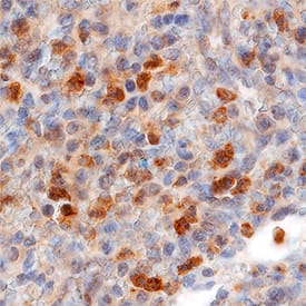
CCR2 was detected in immersion fixed paraffin-embedded sections of human tonsil using Mouse Anti-Human CCR2 Monoclonal Antibody (Catalog # MAB150) at 50 µg/mL overnight at 4 °C. Tissue was stained using the Anti-Mouse HRP-DAB Cell & Tissue Staining Kit (brown; Catalog # CTS002) and counterstained with hematoxylin (blue). Specific staining was localized to lymphocytes. View our protocol for Chromogenic IHC Staining of Paraffin-embedded Tissue Sections.Applications for Human CCR2 Antibody
Application
Recommended Usage
CyTOF-ready
Ready to be labeled using established conjugation methods. No BSA or other carrier proteins that could interfere with conjugation.
Flow Cytometry
0.25 µg/106 cells
Sample: Human peripheral blood monocytes
Sample: Human peripheral blood monocytes
Immunohistochemistry
8-50 µg/mL
Sample: Immersion fixed paraffin-embedded sections of human tonsil
Sample: Immersion fixed paraffin-embedded sections of human tonsil
Reviewed Applications
Read 1 review rated 5 using MAB150 in the following applications:
Formulation, Preparation, and Storage
Purification
Protein A or G purified from ascites
Reconstitution
Reconstitute at 0.5 mg/mL in sterile PBS. For liquid material, refer to CoA for concentration.
Formulation
Lyophilized from a 0.2 μm filtered solution in PBS with Trehalose. *Small pack size (SP) is supplied either lyophilized or as a 0.2 µm filtered solution in PBS.
Shipping
Lyophilized product is shipped at ambient temperature. Liquid small pack size (-SP) is shipped with polar packs. Upon receipt, store immediately at the temperature recommended below.
Stability & Storage
Use a manual defrost freezer and avoid repeated freeze-thaw cycles.
- 12 months from date of receipt, -20 to -70 °C as supplied.
- 1 month, 2 to 8 °C under sterile conditions after reconstitution.
- 6 months, -20 to -70 °C under sterile conditions after reconstitution.
Background: CCR2
Alternate Names
CC-CKR-2, CCR2, CCR2B, CD192, CKR2, CKR2A, CMKBR2, FLJ78302, MCP-1-R
Gene Symbol
CCR2
UniProt
Additional CCR2 Products
Product Documents for Human CCR2 Antibody
Product Specific Notices for Human CCR2 Antibody
For research use only
Loading...
Loading...
Loading...
Loading...
Loading...
