
PD-L1 Antibody (MSVA-711R) - Azide and BSA Free
Novus Biologicals, part of Bio-Techne | Catalog # NBP3-57280
Recombinant Monoclonal Antibody

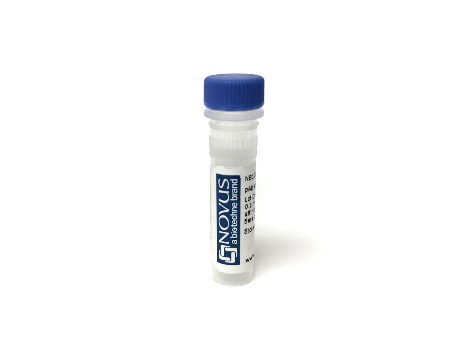
Key Product Details
Species Reactivity
Human
Applications
Immunohistochemistry, Immunohistochemistry-Paraffin
Label
Unconjugated
Antibody Source
Monoclonal Rabbit IgG Clone # MSVA-711R
Format
Azide and BSA Free
Concentration
1 mg/ml
Product Specifications
Immunogen
KLH-conjugated linear peptide corresponding to human PD-L1 Swissprot Q9NZQ7
Localization
Predominantly membranous
Clonality
Monoclonal
Host
Rabbit
Isotype
IgG
Description
This antibody is highly specific and optimized for use in Immunohistochemistry.
It has been validated on TMAs covering 76 normal tissues and numerous cancer tissues.
Positive Control
Postive control:Alveolar macrophages, tonsil crypt epithelium and the placental cytotrophoblast show strong, predominantly membranous positivity. Follicle centre macrophages should show at least moderate intensity staining.
Negative Control
Negitve control:Appendix (negative staining of all epithelial cells)
Applications for PD-L1 Antibody (MSVA-711R) - Azide and BSA Free
Application
Recommended Usage
Immunohistochemistry
Optimal dilutions of this antibody should be experimentally determined.
Immunohistochemistry-Paraffin
Optimal dilutions of this antibody should be experimentally determined.
Application Notes
Manual Protocol: Freshly cut sections should be used (less than 10 days between cutting and staining). Heat-induced antigen retrieval for 5 minutes in an autoclave at 121C in pH 7,8 Target Retrieval Solution buffer. Apply the antibody at a dilution of 1:150 at 37C for 60 minutes. Visualization of bound antibody by the EnVision Kit (Dako, Agilent) according to the manufacturer's directions.
Formulation, Preparation, and Storage
Purification
Protein A or G purified
Format
Azide and BSA Free
Preservative
No Preservative
Concentration
1 mg/ml
Shipping
The product is shipped with polar packs. Upon receipt, store it immediately at the temperature recommended below.
Stability & Storage
Store at 4C short term. Aliquot and store at -20C long term. Avoid freeze-thaw cycles.
Background: PD-L1/B7-H1
PD-L1 binding with receptor PD-1 results in phosphorylation of in the inhibitory tyrosine-based switch motif (ITSM) domain of PD-1, which leads to recruitment of Src homology 2 domain-containing protein tyrosine-phosphatase 2 (SHP-2) and eventual downstream phosphorylation of spleen tyrosine kinase (Syk) and phospholipid inositol-3-kinase (PI3K) (1,3). Under normal conditions, the PD-L1/PD-1 signaling axis helps maintain immune tolerance and prevent destructive immune responses by inhibiting T cell activity such as proliferation, survival, cytokine production, and cytotoxic T lymphocyte (CTL) cytotoxicity (1-3). In the tumor microenvironment (TME), however, the PD-L1/PD-1 signaling axis is hijacked to promote tumor cell survival and limit anti-tumor immune response (1,3). More precisely, tumor cells can escape killing and immune surveillance due to T cell exhaustion and apoptosis (1-3).
Given the role the PD-L1/PD-1 signaling axis plays in tumor cells' ability to evade immune surveillance, it has become a target of several immunotherapeutic agents in recent years (3,5). Antibody immunotherapies that target these inhibitory checkpoint molecules has shown great promise for cancer treatment (3,5). PD-L1 and PD-1 blocking agents have been approved for treatment in a number of cancers including melanoma, non-small cell lung cancer (NSCLC), urothelial carcinoma, and Merkel-cell carcinoma (3,5). In many cancers the expression of PD-L1 in the TME has predictive value for response to blocking agents (3). Pembrolizumab, for example, is a PD-1 inhibitor that has been approved by the FDA as a second-line therapy for treatment of metastatic NSCLC in patients whose tumors express PD-L1 with a Tumor Proportion Score (TPS) greater than 1%, but also for first-line treatment in cases where patients' tumors expression PD-L1 with a TPS greater than 50%) (5). The most promising cancer immunotherapy treatments seem to point to combination therapy with both anti-cancer drugs (e.g. Gefitibin, Metformin, Etoposide) with PD-L1/PD-1 antibody blockade inhibitors (e.g. Atezolizumab, Nivolumab) (6).
References
1. Han, Y., Liu, D., & Li, L. (2020). PD-1/PD-L1 pathway: current researches in cancer. American journal of cancer research, 10(3), 727-742.
2. Jiang, Y., Chen, M., Nie, H., & Yuan, Y. (2019). PD-1 and PD-L1 in cancer immunotherapy: clinical implications and future considerations. Human vaccines & immunotherapeutics, 15(5), 1111-1122. https://doi.org/10.1080/21645515.2019.1571892
3. Sun, C., Mezzadra, R., & Schumacher, T. N. (2018). Regulation and Function of the PD-L1 Checkpoint. Immunity, 48(3), 434-452. https://doi.org/10.1016/j.immuni.2018.03.014
4. Cha, J. H., Chan, L. C., Li, C. W., Hsu, J. L., & Hung, M. C. (2019). Mechanisms Controlling PD-L1 Expression in Cancer. Molecular cell, 76(3), 359-370. https://doi.org/10.1016/j.molcel.2019.09.030
5. Tsoukalas, N., Kiakou, M., Tsapakidis, K., Tolia, M., Aravantinou-Fatorou, E., Baxevanos, P., Kyrgias, G., & Theocharis, S. (2019). PD-1 and PD-L1 as immunotherapy targets and biomarkers in non-small cell lung cancer. Journal of B.U.ON. : official journal of the Balkan Union of Oncology, 24(3), 883-888.
6. Gou, Q., Dong, C., Xu, H., Khan, B., Jin, J., Liu, Q., Shi, J., & Hou, Y. (2020). PD-L1 degradation pathway and immunotherapy for cancer. Cell death & disease, 11(11), 955. https://doi.org/10.1038/s41419-020-03140-2
Long Name
Programmed Death Ligand 1
Alternate Names
B7-H1, B7H1, CD274, PDCD1L1, PDCD1LG1, PDL1
Gene Symbol
CD274
Additional PD-L1/B7-H1 Products
Product Documents for PD-L1 Antibody (MSVA-711R) - Azide and BSA Free
Product Specific Notices for PD-L1 Antibody (MSVA-711R) - Azide and BSA Free
This product is for research use only and is not approved for use in humans or in clinical diagnosis. Primary Antibodies are guaranteed for 1 year from date of receipt.
Loading...
Loading...
Loading...
Loading...