
Mouse Endocan/ESM-1 Antibody
R&D Systems, part of Bio-Techne | Catalog # AF1999


Scientific images may be resized, cropped, or adjusted for brightness or contrast for presentation purposes. These updates are limited to presentation and do not affect the underlying scientific interpretation of the data. Where available, additional source data may be provided upon request.
Conjugate
Catalog #
Key Product Details
Species Reactivity
Validated:
Mouse
Cited:
Mouse, Transgenic Mouse
Applications
Validated:
Immunohistochemistry, Western Blot
Cited:
Immunohistochemistry, Immunohistochemistry-Frozen, Western Blot
Label
Unconjugated
Antibody Source
Polyclonal Goat IgG
Product Specifications
Immunogen
Mouse myeloma cell line NS0-derived recombinant mouse Endocan/ESM-1
Trp20-Arg184
Accession # Q9QYY7
Trp20-Arg184
Accession # Q9QYY7
Specificity
Detects mouse Endocan/ESM-1 in direct ELISAs and Western blots. In direct ELISAs and Western blots, approximately 25% cross-reactivity with recombinant human Endocan is observed.
Clonality
Polyclonal
Host
Goat
Isotype
IgG
Endotoxin Level
<0.10 EU per 1 μg of the antibody by the LAL method.
Scientific Data Images for Mouse Endocan/ESM-1 Antibody
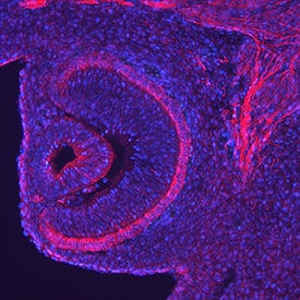
Endocan/ESM‑1 in Mouse Embryo.
Endocan/ESM-1 was detected in perfusion fixed frozen sections of mouse embryo (13 d.p.c.) using Goat Anti-Mouse Endocan/ESM-1 Antigen Affinity-purified Polyclonal Antibody (Catalog # AF1999) at 5 µg/mL overnight at 4 °C. Tissue was stained using the NorthernLights™ 557-conjugated Anti-Goat IgG Secondary Antibody (red; Catalog # NL001) and counterstained with DAPI (blue). Specific staining was localized to the retina. View our protocol for Fluorescent IHC Staining of Frozen Tissue Sections.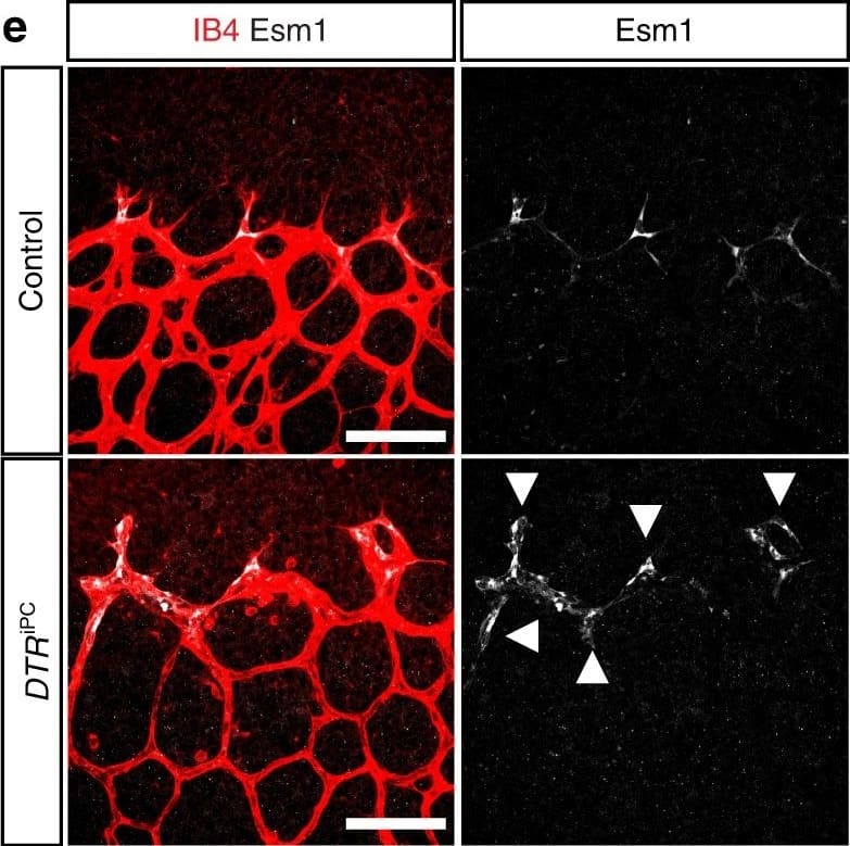
Detection of Mouse Endocan/ESM-1 by Immunocytochemistry/Immunofluorescence
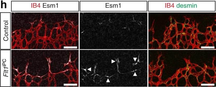
Endothelial changes after pericyte depletion. a–f Maximum intensity projection of confocal images from control and DTRiPC P6 retinas stained for IB4 (red) in combination with VEGF-A a, VEGFR2 b, VEGFR3 c, Tie2 d, Esm1 e, and Dll4 f (all in white), as indicated. Note local increase of VEGFR2, VEGFR3, and Esm1 (arrowheads in b, c, e) but not Tie2 or VEGF-A at the edge of the vessel plexus. Dll4 expression in DTRiPC sprouts is increased in some regions (arrowheads) but absent in others (arrows). Scale bar, 100 µm. g–j Quantitation of VEGF-A immunosignals area and intensity g, signal intensity for VEGFR2 h and VEGFR3 i and proportion of Esm1+ area with respect to vascular area j in the P6 control and DTRiPC angiogenic front. Error bars, s.e.m. p-values, Student’s t-test Image collected and cropped by CiteAb from the following publication (https://pubmed.ncbi.nlm.nih.gov/29146905), licensed under a CC-BY license. Not internally tested by R&D Systems.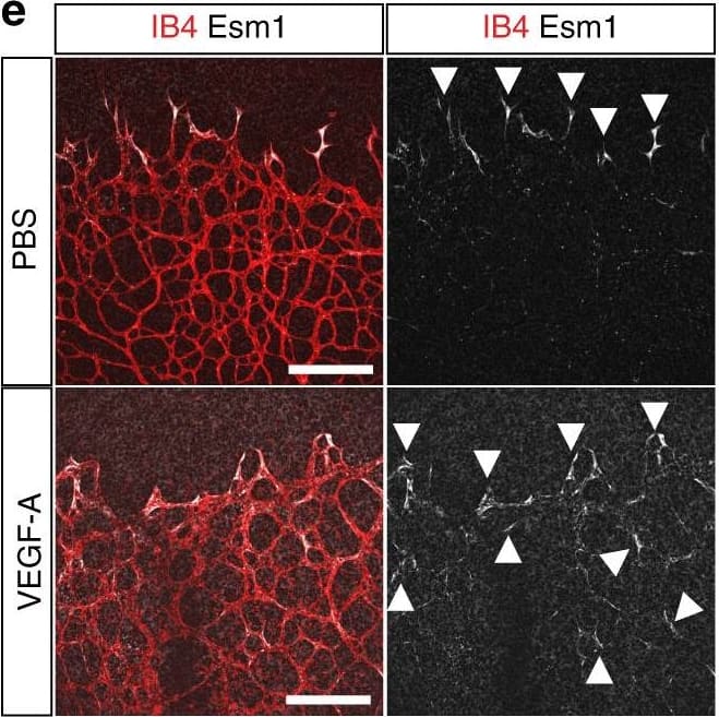
Detection of Mouse Endocan/ESM-1 by Immunocytochemistry/Immunofluorescence
Vascular alterations after intraocular VEGF-A injection. a Morphology of IB4-stained P6 wild-type retinal vessels at 4 h after administration of human VEGF-A165 (0.5 µl at a concentration of 5 μg μl−1). Note blunt appearance of the vessel front after VEGF-A injection but not for vehicle (PBS) control. Scale bar, 200 µm. b Quantitation of sprouts and filopodia at the front of the P6 vessel plexus after injection of VEGF-A165 or vehicle control. Error bars, s.e.m. p-values, Student’s t-test. c PDGFR beta+ (green) pericytes are unaffected by short-term VEGF-A administration, whereas VEGFR2 immunosignals (white) are increased in IB4+ (red) ECs (arrowheads). Images shown correspond to insets in a. Scale bar, 100 µm. d Quantitation of VEGFR2 immunosignals intensity in the peripheral plexus of P6 retinas after injection of VEGF-A165 or vehicle control. Error bars, s.e.m. p-values, Student’s t-test. e Confocal images showing increased Esm1 immunostaining (white) in IB4+ (red) ECs in the peripheral plexus (arrowheads) after VEGF-A injection in P6 pups. Scale bar, 200 µm. f VEGF-A165 injection-mediated increase of Esm1 immunosignals (normalized to IB4+ EC area) in the peripheral capillary plexus but not at the edge of the angiogenic front in comparison to PBS-injected controls at P6. Error bars, s.e.m. p-values, Student’s t-test. NS, not statistically significant. g Short-term VEGF-A165 administration leads to clustering of Erg1+ (green) and IB4+ (red) ECs, as indicated, in thick sprout-like structures of P6 retinas. Panels in the center and on the right (scale bar, 20 µm) show higher magnification of the insets on the left (scale bar, 100 µm). Dashed lines in panels on the right outline IB4+ vessels. h Quantitation of EC density in the leading front vessel and emerging sprouts of the P6 angiogenic front after injection of VEGF-A165 or vehicle control. Error bars, s.e.m. p-values, Student’s t-test Image collected and cropped by CiteAb from the following publication (https://pubmeApplications for Mouse Endocan/ESM-1 Antibody
Application
Recommended Usage
Immunohistochemistry
5-15 µg/mL
Sample:
Sample:
Perfusion fixed frozen sections of mouse embryo (13 d.p.c.)
Western Blot
0.1 µg/mL
Sample: Recombinant Mouse Endocan/ESM-1 (Catalog # 1999-EC)
Sample: Recombinant Mouse Endocan/ESM-1 (Catalog # 1999-EC)
Formulation, Preparation, and Storage
Purification
Antigen Affinity-purified
Reconstitution
Reconstitute at 0.2 mg/mL in sterile PBS. For liquid material, refer to CoA for concentration.
Formulation
Lyophilized from a 0.2 μm filtered solution in PBS with Trehalose. See Certificate of Analysis for details.
*Small pack size (-SP) is supplied either lyophilized or as a 0.2 µm filtered solution in PBS.
*Small pack size (-SP) is supplied either lyophilized or as a 0.2 µm filtered solution in PBS.
Shipping
Lyophilized product is shipped at ambient temperature. Liquid small pack size (-SP) is shipped with polar packs. Upon receipt, store immediately at the temperature recommended below.
Stability & Storage
Use a manual defrost freezer and avoid repeated freeze-thaw cycles.
- 12 months from date of receipt, -20 to -70 °C as supplied.
- 1 month, 2 to 8 °C under sterile conditions after reconstitution.
- 6 months, -20 to -70 °C under sterile conditions after reconstitution.
Background: Endocan/ESM-1
Endocan (endothelial cell proteoglycan; also known as endothelial-cell specific molecule-1 [ESM-1]), is a 50 kDa, monomeric, secreted, cysteine-rich proteoglycan identified initially in endothelial cells of the kidney and lung (1). Mouse Endocan is synthesized as a 184 amino acid (aa) precursor that contains a 21 aa signal sequence and a 20 kDa, 163 aa mature region (2). The N-terminal 2/3 of the molecule contains 18 cysteine residues and there are no potential N-linked glycosylation sites. Based on human Endocan, there are at least two potential O-linked glycosylation sites, one of which will likely be utilized on Ser at position # 136 of the mature molecule (3). The posttranslational modification is approximately 30 kDa in size. It consists of a single dermatan sulfate chain that contains 4-O sulfated N-acetyl galactosamine with alpha-iduronate. This chain is suggested to bind HGF and contribute to HGF mitogenic activity (4). Mature mouse Endocan is 96% and 74% aa identical to rat and human Endocan, respectively. In human, there is a potential for an alternate splice variant. It shows a deletion of aa’s 82 through 131, a range which would not remove the dermatan sulfate attachment site (4). It is not known if such a splice form exists in mouse. Endocan is expressed by endothelial cells, adipocytes, bronchial epithelium and distal renal tubular epithelium (1, 5, 6). It is upregulated by TNF-alpha and VEGF, (1, 7) and is known to bind to LFA-1 ( alphaL beta2) on the surface of PBMCs, blocking LFA-1 interaction with ICAM-1 (8). Normal circulating levels of Endocan are approximately 1 ng/mL (6).
References
- Lassalle, P. et al. (1996) J. Biol. Chem. 271:20458.
- Lassalle, P. (1999) Genbank Accession #: Q9QYY7.
- Bechard, D. et al. (2001) J. Biol. Chem. 276:48341.
- Aitkenhead, M. et al. (2002) Microvasc. Res. 63:159.
- Wellner, M. et al. (2003) Horm. Metab. Res. 35:217
- Bechard, D. et al. (2000) J. Vasc. Res. 37:417.
- Tsai, J.C. et al. (2002) J. Vasc. Res. 39:148.
Alternate Names
ESM-1, ESM1, IGFBP-rp6
Gene Symbol
ESM1
UniProt
Additional Endocan/ESM-1 Products
Product Documents for Mouse Endocan/ESM-1 Antibody
Product Specific Notices for Mouse Endocan/ESM-1 Antibody
For research use only
Loading...
Loading...
Loading...
Loading...
Loading...