Mouse CD3 Antibody
R&D Systems, part of Bio-Techne | Catalog # MAB4841

Conjugate
Catalog #
Key Product Details
Validated by
Knockout/Knockdown
Species Reactivity
Validated:
Mouse
Cited:
Human, Mouse, Transgenic Mouse
Applications
Validated:
Immunohistochemistry, Flow Cytometry, Immunocytochemistry, Immunoprecipitation, Complement-dependent Cytotoxicity, T Cell Stimulation, CyTOF-ready
Cited:
Immunohistochemistry, Immunohistochemistry-Paraffin, Neutralization, Flow Cytometry, Immunofluorescence, Immunocytochemistry, Activation, Bioassay, ELISA Capture, Functional Assay
Label
Unconjugated
Antibody Source
Monoclonal Rat IgG2B Clone # 17A2
Product Specifications
Immunogen
T cell hybridoma D1
Specificity
Reacts with mouse TCR-associated CD3 complex that occurs on thymocytes and mature T cells.
Clonality
Monoclonal
Host
Rat
Isotype
IgG2B
Endotoxin Level
<0.10 EU per 1 μg of the antibody by the LAL method.
Scientific Data Images for Mouse CD3 Antibody
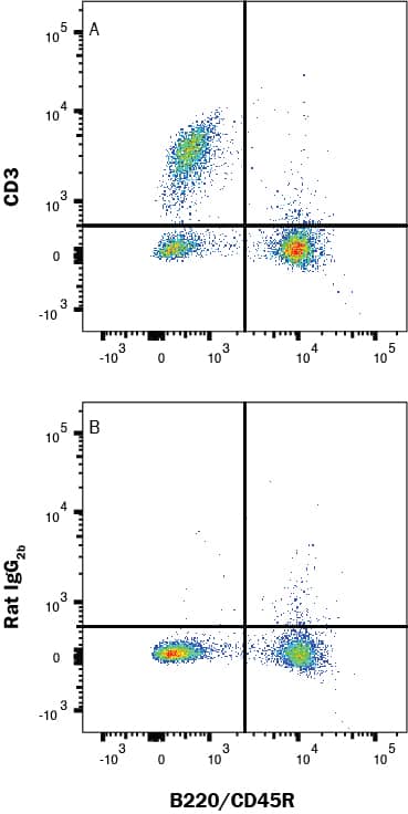
Detection of CD3 in mouse splenocytes by Flow Cytometry
Mouse splenocytes were stained with Rat Anti-Mouse B220/CD45R PE-conjugated Monoclonal Antibody (Catalog # FAB1217P) and either (A) Rat Anti-Mouse CD3 Monoclonal Antibody (Catalog # MAB4841) or (B) isotype control antibody (Catalog # MAB0061) followed by Allophycocyanin-conjugated Anti-Rat IgG Secondary Antibody (Catalog # F0113). View our protocol for Staining Membrane-associated Proteins.CD3 in Mouse Splenocytes.
CD3 was detected in immersion fixed mouse splenocytes using Rat Anti-Mouse CD3 Monoclonal Antibody (Catalog # MAB4841) at 3 µg/mL for 3 hours at room temperature. Cells were stained using the NorthernLights™ 557-conjugated Anti-Rat IgG Secondary Antibody (red; NL013) and counterstained with DAPI (blue). Specific staining was localized to plasma membrane. View our protocol for Fluorescent ICC Staining of Non-adherent Cells.CD3 in Mouse Brain.
CD3 was detected in perfusion fixed HSV-1 infected frozen sections of mouse brain (perivascular cuffing) using 10 µg/mL Rat Anti-Mouse CD3 Monoclonal Antibody (Catalog # MAB4841) overnight at 4 °C. Tissue was stained using the NorthernLights™ 493-conjugated Anti-Rat IgG Secondary Antibody (green; NL015) and counterstained with DAPI (blue). View our protocol for Fluorescent IHC Staining of Frozen Tissue Sections.Applications for Mouse CD3 Antibody
Application
Recommended Usage
Complement-dependent Cytotoxicity
Miescher, G.C. et al. (1989) Immunol. Lett. 23:113 and Wu, L. et al. (1991) J. Exp. Med. 174:1617.
CyTOF-ready
Ready to be labeled using established conjugation methods. No BSA or other carrier proteins that could interfere with conjugation.
Flow Cytometry
0.25 µg/106 cells
Sample: B220+ Mouse splenocytes
Sample: B220+ Mouse splenocytes
Immunocytochemistry
3-25 µg/mL
Sample: Immersion fixed mouse splenocytes
Sample: Immersion fixed mouse splenocytes
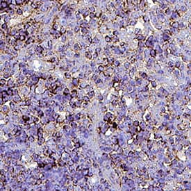
Immunohistochemistry
8-25 µg/mL
Sample: Perfusion fixed HSV-1 infected frozen sections of mouse brain (perivascular cuffing) and perfusion fixed paraffin-embedded sections of Mouse Thymus
Sample: Perfusion fixed HSV-1 infected frozen sections of mouse brain (perivascular cuffing) and perfusion fixed paraffin-embedded sections of Mouse Thymus
Immunoprecipitation
Miescher, G.C. et al. (1989) Immunol. Lett. 23:113.
T Cell Stimulation
Miescher, G.C. et al. (1989) Immunol. Lett. 23:113.
Reviewed Applications
Read 5 reviews rated 5 using MAB4841 in the following applications:
Formulation, Preparation, and Storage
Purification
Protein A or G purified from hybridoma culture supernatant
Reconstitution
Reconstitute at 0.5 mg/mL in sterile PBS. For liquid material, refer to CoA for concentration.
Formulation
Lyophilized from a 0.2 μm filtered solution in PBS with Trehalose. See Certificate of Analysis for details.
*Small pack size (-SP) is supplied either lyophilized or as a 0.2 µm filtered solution in PBS.
*Small pack size (-SP) is supplied either lyophilized or as a 0.2 µm filtered solution in PBS.
Shipping
Lyophilized product is shipped at ambient temperature. Liquid small pack size (-SP) is shipped with polar packs. Upon receipt, store immediately at the temperature recommended below.
Stability & Storage
Use a manual defrost freezer and avoid repeated freeze-thaw cycles.
- 12 months from date of receipt, -20 to -70 °C as supplied.
- 1 month, 2 to 8 °C under sterile conditions after reconstitution.
- 6 months, -20 to -70 °C under sterile conditions after reconstitution.
Background: CD3
References
- Miescher, G.C. et al. (1989) Immunol. Lett. 23:113.
Alternate Names
CD_antigen: CD3e, CD3 antigen, delta subunit, CD3d antigen, CD3d antigen, delta polypeptide (TiT3 complex), CD3d molecule, delta (CD3-TCR complex), CD3-DELTA, CD3e, CD3e antigen, CD3e antigen, epsilon polypeptide (TiT3 complex), CD3e molecule, epsilon (CD3-TCR complex), CD3-epsilon, CD3G, CD3g antigen, CD3g antigen, gamma polypeptide (TiT3 complex), CD3g molecule, epsilon (CD3-TCR complex), CD3g molecule, gamma (CD3-TCR complex), CD3-GAMMA, FLJ17620, FLJ17664, FLJ18683, FLJ79544, FLJ94613, IMD18, MGC138597, T3DOKT3, delta chain, T3E, T-cell antigen receptor complex, epsilon subunit of T3, T-cell receptor T3 delta chain, T-cell surface antigen T3/Leu-4 epsilon chain, T-cell surface glycoprotein CD3 delta chain, T-cell surface glycoprotein CD3 epsilon chain, TCRE
Gene Symbol
CD3E
Additional CD3 Products
Product Documents for Mouse CD3 Antibody
Product Specific Notices for Mouse CD3 Antibody
For research use only
Loading...
Loading...
Loading...
Loading...
Loading...


