
Mouse Caspase-11/Caspase-4 Antibody
R&D Systems, part of Bio-Techne | Catalog # MAB8648
Recombinant Monoclonal Antibody.


Scientific images may be resized, cropped, or adjusted for brightness or contrast for presentation purposes. These updates are limited to presentation and do not affect the underlying scientific interpretation of the data. Where available, additional source data may be provided upon request.
Conjugate
Catalog #
Key Product Details
Validated by
Biological Validation
Species Reactivity
Mouse
Applications
Western Blot
Label
Unconjugated
Antibody Source
Recombinant Monoclonal Rabbit IgG Clone # 1172C
Product Specifications
Immunogen
HEK293 human embryonic kidney cell line transfected with mouse Caspase-11/Caspase-4
Accession # P70343
Accession # P70343
Specificity
Detects mouse Caspase-11/Caspase-4 in Western blots.
Clonality
Monoclonal
Host
Rabbit
Isotype
IgG
Scientific Data Images for Mouse Caspase-11/Caspase-4 Antibody
Detection of Mouse Caspase-11/Caspase-4 by Western Blot.
Western blot shows lysates of mouse splenocytes untreated (-) or treated (+) with LPS. PVDF membrane was probed with 1 µg/mL of Rabbit Anti-Mouse Caspase-11/Caspase-4 Monoclonal Antibody (Catalog # MAB8648) followed by HRP-conjugated Anti-Rabbit IgG Secondary Antibody (Catalog # HAF008). Specific bands were detected for Caspase-11/Caspase-4 at approximately 36, 44, and 46 kDa (as indicated). This experiment was conducted under reducing conditions and using Immunoblot Buffer Group 1.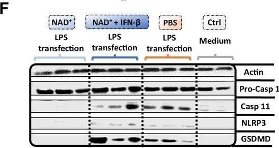
Detection of Mouse Caspase-11/Caspase-4 by Western Blot
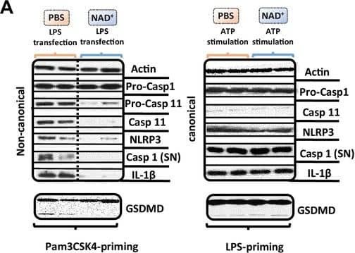
Pro-casp-1, casp-11, NLRP3, gasdermin D (GSDMD) expression were assessed by western blot. Statistical significance was determined by using Student’s t-test or one-way ANOVA. Asterisks indicate p-values *=p<0.05, **=p<0.01, and ***=p<0.001, only significant values are shown. All data depicted in this figure are provided as source data. Image collected and cropped by CiteAb from the following open publication (https://elifesciences.org/articles/88686), licensed under a CC-BY license. Not internally tested by R&D Systems.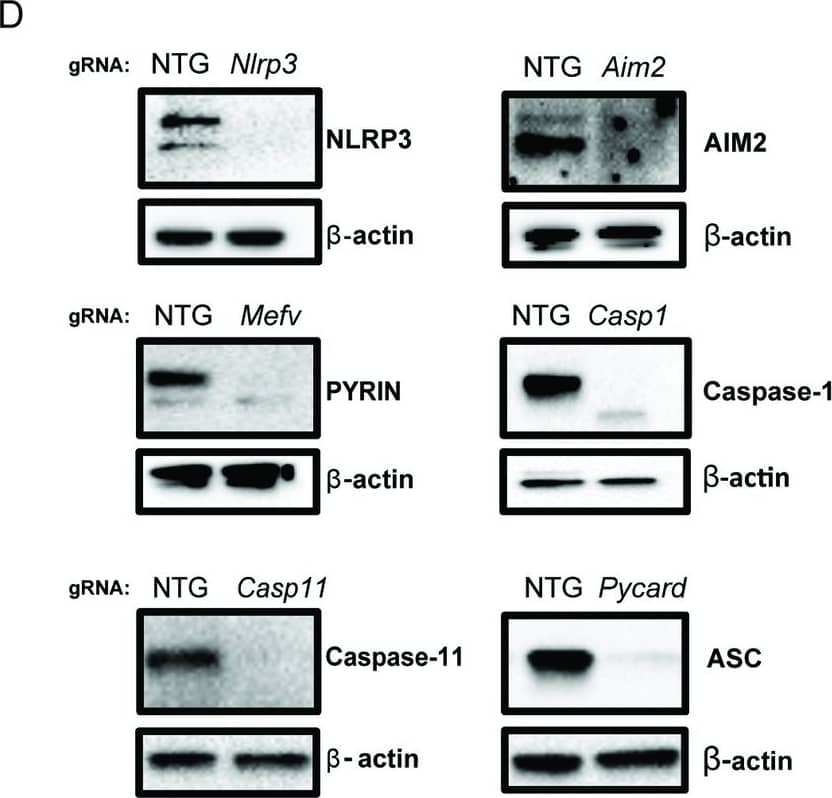
Detection of Mouse Caspase-11/Caspase-4 by Western Blot
DENV NS1-induced inflammasome activation is NLPR3-independent.(A) WT and Nlrp3-/- BMDMs were primed with PAM3CSK4 (1μg/mL) for 17h and then treated with DENV2 NS1 at indicated concentrations, nigericin (5μM), or medium (PAM only). IL-1 beta levels in supernatant 2h (nigericin) or 24h (NS1 and PAM only) were measured by ELISA. Statistical significance was determined using two-way ANOVA with Holm-Sidak’s multiple comparisons test. (B) Representative Western blots of cell lysates from WT and Nlrp3-/- BMDMs after priming with PAM3CSK4 (1μg/mL) for 17h and treatment with DENV2 NS1 (10 or 5 μg/mL), treatment with nigericin (5μM), or no treatment for 24h. (C) BMDMs were primed with PAM3CSK4 (1μg/mL) for 17h and then pre-treated with MCC950 at the indicated concentrations before addition of DENV2 NS1 (10μg/mL), nigericin (5μM), or medium (Inhibitor only). IL-1 beta levels in the supernatant after 2h (Nigericin) or 24h (NS1 and PAM only) were measured by ELISA. (D) Representative Western blots of cell lysates from BMDMs nucleofected with Cas9-gRNA ribonuclear protein complexes to knock out the indicated genes. Two gRNAs per gene were used per nucleofection. NTG = non-targeting guide. (E) Knockout BMDMs from (D) were primed with PAM3CSK4 (1μg/mL) for 17h and treated with DENV2 NS1 (10μg/mL) or left untreated for 48h. Statistical significance was determined using two-way ANOVA followed by with Holm-Sidak’s multiple comparisons test. The data are shown as the mean ± SD of 3 biological replicates (A,C), a representative image taken from 2 biological replicates (B,D), or data pooled from 8 independent experiments with at least 3 biological replicates per guide (E). *p<0.05, **p<0.01, *** p< 0.001, ****p<0.0001, ns (not significant), p> 0.05. Image collected and cropped by CiteAb from the following open publication (https://dx.plos.org/10.1371/journal.ppat.1012167), licensed under a CC-BY license. Not internally tested by R&D Systems.Applications for Mouse Caspase-11/Caspase-4 Antibody
Application
Recommended Usage
Western Blot
1 µg/mL
Sample: Mouse splenocytes treated with LPS
Sample: Mouse splenocytes treated with LPS
Reviewed Applications
Read 1 review rated 4 using MAB8648 in the following applications:
Formulation, Preparation, and Storage
Purification
Protein A or G purified from cell culture supernatant
Reconstitution
Reconstitute at 0.5 mg/mL in sterile PBS. For liquid material, refer to CoA for concentration.
Formulation
Lyophilized from a 0.2 μm filtered solution in PBS with Trehalose. *Small pack size (SP) is supplied either lyophilized or as a 0.2 µm filtered solution in PBS.
Shipping
Lyophilized product is shipped at ambient temperature. Liquid small pack size (-SP) is shipped with polar packs. Upon receipt, store immediately at the temperature recommended below.
Stability & Storage
Use a manual defrost freezer and avoid repeated freeze-thaw cycles.
- 12 months from date of receipt, -20 to -70 °C as supplied.
- 1 month, 2 to 8 °C under sterile conditions after reconstitution.
- 6 months, -20 to -70 °C under sterile conditions after reconstitution.
Background: Caspase-11/Caspase-4
Alternate Names
CASP11, Caspl, Ich-3
Gene Symbol
CASP4
UniProt
Additional Caspase-11/Caspase-4 Products
Product Documents for Mouse Caspase-11/Caspase-4 Antibody
Product Specific Notices for Mouse Caspase-11/Caspase-4 Antibody
For research use only
Loading...
Loading...
Loading...
Loading...
Loading...