
Human VEGFR2/KDR/Flk-1 Antibody
R&D Systems, part of Bio-Techne | Catalog # MAB3572


Scientific images may be resized, cropped, or adjusted for brightness or contrast for presentation purposes. These updates are limited to presentation and do not affect the underlying scientific interpretation of the data. Where available, additional source data may be provided upon request.
Conjugate
Catalog #
Key Product Details
Species Reactivity
Validated:
Human
Cited:
Human, Mouse, Primate - Macaca mulatta (Rhesus Macaque)
Applications
Validated:
Neutralization, Flow Cytometry, CyTOF-ready
Cited:
Neutralization, Flow Cytometry
Label
Unconjugated
Antibody Source
Monoclonal Mouse IgG1 Clone # 89106
Product Specifications
Immunogen
S. frugiperda insect ovarian cell line Sf 21-derived recombinant human VEGFR2/KDR/Flk-1
Ala20-Glu764
Accession # P35968
Ala20-Glu764
Accession # P35968
Specificity
Detects human VEGFR2/KDR/Flk-1 in direct ELISAs. In direct ELISAs, no cross-reactivity with recombinant human (rh) VEGFR1, rhVEGFR3 or recombinant mouse VEGFR2 is observed.
Clonality
Monoclonal
Host
Mouse
Isotype
IgG1
Endotoxin Level
<0.10 EU per 1 μg of the antibody by the LAL method.
Scientific Data Images for Human VEGFR2/KDR/Flk-1 Antibody
Detection of VEGFR2/KDR/Flk‑1 in HUVEC Human Cells by Flow Cytometry.
HUVEC human umbilical vein endothelial cells were stained with Mouse Anti-Human VEGFR2/KDR/Flk-1 Monoclonal Antibody (Catalog # MAB3572, filled histogram) or isotype control antibody (Catalog # MAB002, open histogram), followed by Phycoerythrin-conjugated Anti-Mouse IgG Secondary Antibody (Catalog # F0102B).
VEGFR2/KDR Inhibition of VEGF-dependent Cell Proliferation and Neutralization by Human VEGFR2/KDR Antibody.
Recombinant Human VEGFR2/KDR Fc Chimera (Catalog # 357-KD) inhibits Recombinant Human VEGF165(Catalog # 293-VE) induced proliferation in HUVEC human umbilical vein endothelial cells in a dose-dependent manner (orange line). Inhibition of Recombinant Human VEGF165(10 ng/mL) activity elicited by Recombinant Human VEGFR2/KDR Fc Chimera (50 ng/mL) is neutralized (green line) by increasing concentrations of Mouse Anti-Human VEGFR2/KDR Monoclonal Antibody (Catalog # MAB3572). The ND50 is typically 10-50 ng/mL.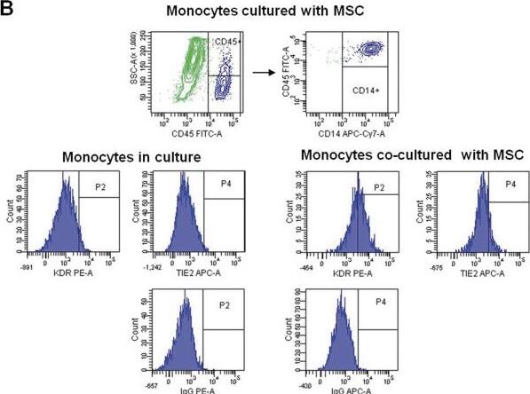
Detection of VEGFR2/KDR/Flk-1 by Flow Cytometry
Induction of endothelial markers in circulating CD14+ cells. CD14+ monocytes were isolated from adult PB, cultured in EGM-2 MV with or without MSCs, and then analyzed with flow cytometry. Dot plots from freshly isolated CD14+ cells (A) and after 4 days of co-culture with adipose-derived MSCs (green) (B) are shown. Flow-cytometry histograms in panels A and B show the expression of KDR and Tie2 in gated CD14+ cells. Isotype-matched controls are given. Image collected and cropped by CiteAb from the following open publication (https://pubmed.ncbi.nlm.nih.gov/24731246), licensed under a CC-BY license. Not internally tested by R&D Systems.Applications for Human VEGFR2/KDR/Flk-1 Antibody
Application
Recommended Usage
CyTOF-ready
Ready to be labeled using established conjugation methods. No BSA or other carrier proteins that could interfere with conjugation.
Flow Cytometry
0.25 µg/106 cells
Sample: HUVEC human umbilical vein endothelial cells
Sample: HUVEC human umbilical vein endothelial cells
Neutralization
Measured by its ability to neutralize VEGFR2/KDR/Flk‑1-mediated inhibition of proliferation in HUVEC human umbilical vein endothelial cells. The Neutralization Dose (ND50) is typically 10-50 ng/mL in the presence of 50 ng/mL Recombinant Human VEGFR2/KDR/Flk‑1 Fc Chimera and 10 ng/mL Recombinant Human VEGF165.
Reviewed Applications
Read 2 reviews rated 5 using MAB3572 in the following applications:
Formulation, Preparation, and Storage
Purification
Protein A or G purified from hybridoma culture supernatant
Reconstitution
Reconstitute at 0.5 mg/mL in sterile PBS. For liquid material, refer to CoA for concentration.
Formulation
Lyophilized from a 0.2 μm filtered solution in PBS with Trehalose. *Small pack size (SP) is supplied either lyophilized or as a 0.2 µm filtered solution in PBS.
Shipping
Lyophilized product is shipped at ambient temperature. Liquid small pack size (-SP) is shipped with polar packs. Upon receipt, store immediately at the temperature recommended below.
Stability & Storage
Use a manual defrost freezer and avoid repeated freeze-thaw cycles.
- 12 months from date of receipt, -20 to -70 °C as supplied.
- 1 month, 2 to 8 °C under sterile conditions after reconstitution.
- 6 months, -20 to -70 °C under sterile conditions after reconstitution.
Background: VEGFR2/KDR/Flk-1
VEGFR2 (KDR/Flk-1), VEGFR1 (Flt-1) and VEGFR3 (Flt-4) belong to the class III subfamily of receptor tyrosine kinases (RTKs). All three receptors contain seven immunoglobulin-like repeats in their extracellular domains and kinase insert domains in their intracellular regions. The expression of VEGFR1, 2, and 3 is almost exclusively restricted to the endothelial cells. These receptors are likely to play essential roles in vasculogenesis and angiogenesis. Mature VEGFR2 is composed of a 745 aa extracellular domain, a 25 aa transmembrane domain and a 567 aa cytoplasmic domain. In contrast to VEGFR1 which binds both PlGF and VEGF with high affinity, VEGFR2 binds VEGF but not PlGF with high affinity. The recombinant soluble VEGFR2/Fc chimera binds VEGF with high affinity and is a potent VEGF antagonist.
References
- Ferra, N. and R. Davis-Smyth (1997) Endocrine Reviews 18:4.
Long Name
Vascular Endothelial Growth Factor Receptor 2
Alternate Names
CD309, Flk-1, Flk1, KDR, KRD1, Ly73, VEGF R2
Gene Symbol
KDR
UniProt
Additional VEGFR2/KDR/Flk-1 Products
Product Documents for Human VEGFR2/KDR/Flk-1 Antibody
Product Specific Notices for Human VEGFR2/KDR/Flk-1 Antibody
For research use only
Loading...
Loading...
Loading...
Loading...
Loading...
Loading...