
Human Phospho-Histone H2AX (S139) Antibody
R&D Systems, part of Bio-Techne | Catalog # MAB2288
Recombinant Monoclonal Antibody.

Scientific images may be resized, cropped, or adjusted for brightness or contrast for presentation purposes. These updates are limited to presentation and do not affect the underlying scientific interpretation of the data. Where available, additional source data may be provided upon request.
Conjugate
Catalog #
Key Product Details
Validated by
Biological Validation
Species Reactivity
Validated:
Human
Cited:
Mouse
Applications
Validated:
Western Blot, Intracellular Staining by Flow Cytometry, CyTOF-ready
Cited:
Immunohistochemistry
Label
Unconjugated
Antibody Source
Recombinant Monoclonal Rabbit IgG Clone # 2207D
Product Specifications
Immunogen
Phospho-peptide containing human Histone H2AX S139 site
Accession # P16104
Accession # P16104
Specificity
Detects human Histone H2AX when phosphorylated at S139 in Western blots.
Clonality
Monoclonal
Host
Rabbit
Isotype
IgG
Scientific Data Images for Human Phospho-Histone H2AX (S139) Antibody
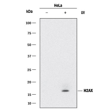
Detection of Human Histone H2AX by Western Blot.
Western blot shows lysates of HeLa human cervical epithelial carcinoma cell line untreated (-) or treated (+) with 20 mJ/cm2 ultraviolet light (UV) followed by a 30 minute recover. PVDF membrane was probed with 0.25 µg/mL of Rabbit Anti-Human Phospho-Histone H2AX (S139) Monoclonal Antibody (Catalog # MAB2288) followed by HRP-conjugated Anti-Rabbit IgG Secondary Antibody (HAF008). A specific band was detected for Histone H2AX at approximately 17 kDa (as indicated). This experiment was conducted under reducing conditions and using Immunoblot Buffer Group 1.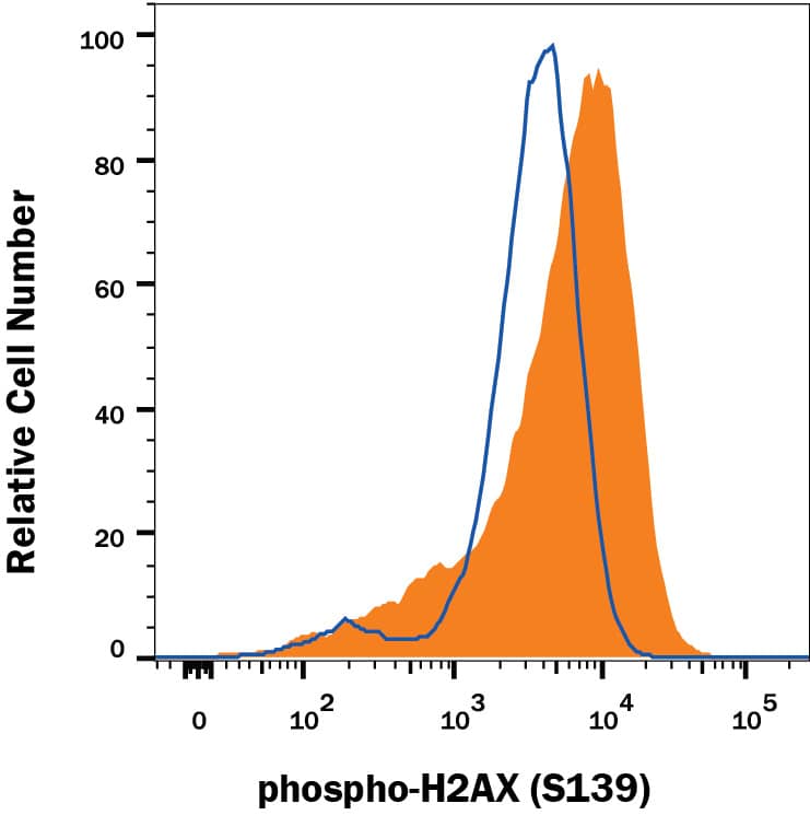
Detection of Human Phospho-Histone H2AX (S139) in Camptothecin-treated Jurkat cells by Flow Cytometry.
Naïve Jurkat cells (open histogram) or treated with 1 µM Camptothecin (1100, filled histogram) overnight were stained with Rabbit Anti-Human Phospho-Histone H2AX (S139) Monoclonal Antibody (Catalog # MAB2288), followed by PE-conjugated Anti-Rabbit IgG Secondary Antibody (F0110). To facilitate intracellular staining, cells were fixed with Flow Cytometry Fixation Buffer (FC004) and permeabilized with 90% methanol. View our protocol for Staining Intracellular Molecules.Applications for Human Phospho-Histone H2AX (S139) Antibody
Application
Recommended Usage
CyTOF-ready
Ready to be labeled using established conjugation methods. No BSA or other carrier proteins that could interfere with conjugation.
Intracellular Staining by Flow Cytometry
0.25 µg/106 cells
Sample: Camptothecin-treated Jurkat cells, fixed with Flow Cytometry Fixation Buffer (Catalog# FC004) and permeabilized with 90% methanol
Sample: Camptothecin-treated Jurkat cells, fixed with Flow Cytometry Fixation Buffer (Catalog# FC004) and permeabilized with 90% methanol
Western Blot
0.25 µg/mL
Sample: HeLa human cervical epithelial carcinoma cell line treated with ultraviolet light (UV)
Sample: HeLa human cervical epithelial carcinoma cell line treated with ultraviolet light (UV)
Reviewed Applications
Read 1 review rated 5 using MAB2288 in the following applications:
Formulation, Preparation, and Storage
Purification
Protein A or G purified from cell culture supernatant
Reconstitution
Reconstitute at 0.5 mg/mL in sterile PBS. For liquid material, refer to CoA for concentration.
Formulation
Lyophilized from a 0.2 μm filtered solution in PBS with Trehalose. *Small pack size (SP) is supplied either lyophilized or as a 0.2 µm filtered solution in PBS.
Shipping
Lyophilized product is shipped at ambient temperature. Liquid small pack size (-SP) is shipped with polar packs. Upon receipt, store immediately at the temperature recommended below.
Stability & Storage
Use a manual defrost freezer and avoid repeated freeze-thaw cycles.
- 12 months from date of receipt, -20 to -70 °C as supplied.
- 1 month, 2 to 8 °C under sterile conditions after reconstitution.
- 6 months, -20 to -70 °C under sterile conditions after reconstitution.
Background: Histone H2AX
Histone H2AX is a core histone protein that is phosphorylated at S139 in cells exposed to DNA double-strand break-inducing agents, such as ionizing radiation. The S139 phosphorylated H2AX, termed gamma-H2AX, marks the site of DNA double-strand breaks and serves to recruit cell cycle checkpoint and DNA repair factors to the site of damage.
Additional Histone H2AX Products
Product Documents for Human Phospho-Histone H2AX (S139) Antibody
Product Specific Notices for Human Phospho-Histone H2AX (S139) Antibody
For research use only
Loading...
Loading...
Loading...
Loading...
Loading...
Loading...