
Human/Mouse/Rat VAMP-2 Antibody
R&D Systems, part of Bio-Techne | Catalog # AF5136


Scientific images may be resized, cropped, or adjusted for brightness or contrast for presentation purposes. These updates are limited to presentation and do not affect the underlying scientific interpretation of the data. Where available, additional source data may be provided upon request.
Conjugate
Catalog #
Key Product Details
Species Reactivity
Validated:
Human, Mouse, Rat
Cited:
Transgenic Mouse
Applications
Validated:
Immunohistochemistry, Western Blot
Cited:
Western Blot, Immunocytochemistry
Label
Unconjugated
Antibody Source
Polyclonal Goat IgG
Product Specifications
Immunogen
E. coli-derived recombinant human VAMP-2
Met1-Lys94
Accession # P63027
Met1-Lys94
Accession # P63027
Specificity
Detects human, mouse, and rat VAMP-2 in direct ELISAs and Western blots. In direct ELISAs, less than 10% cross-reactivity with recombinant human (rh) VAMP-1 is observed and less than 2% cross-reactivity with rhVAMP-3 and rhVAMP-7 is observed.
Clonality
Polyclonal
Host
Goat
Isotype
IgG
Scientific Data Images for Human/Mouse/Rat VAMP-2 Antibody
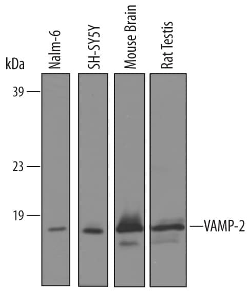
Detection of Human, Mouse, and Rat VAMP‑2 by Western Blot.
Western blot shows lysates of Nalm-6 human Pre-B acute lymphocytic leukemia cell line, SH-SY5Y human neuroblastoma cell line, mouse brain tissue, and rat testis tissue. PVDF membrane was probed with 1 µg/mL of Goat Anti-Human/Mouse/Rat VAMP-2 Antigen Affinity-purified Polyclonal Antibody (Catalog # AF5136) followed by HRP-conjugated Anti-Goat IgG Secondary Antibody (Catalog # HAF019). A specific band was detected for VAMP-2 at approximately 13 kDa (as indicated). This experiment was conducted under reducing conditions and using Immunoblot Buffer Group 8.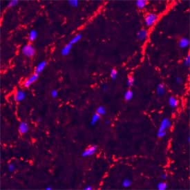
VAMP-2 in Rat Spinal Cord.
Vesicle-Associate Membrane Protein 2 (VAMP-2) was detected in perfusion-fixed frozen sections of rat spinal cord using a Goat Anti-Human/Mouse/Rat VAMP-2 Antigen Affinity-purified Polyclonal Antibody (Catalog # AF5136). The cells were stained with the NorthernLights 557-conjugated Donkey Anti-Goat IgG Affinity-purified Secondary Antibody (red; Catalog # NL001). The cell nuclei were counterstained with DAPI (blue). VAMP-2 immunoreactivity was localized to synaptic vesicles. View our protocol for Fluorescent IHC Staining of Frozen Tissue Sections.
Detection of VAMP-2 by Western Blot
Stx2 deletion enhances exocytosis by facilitating trans-SNARE complex assembly and cis-SNARE complex disassembly.a Stx2 deletion promotes the formation of profusion trans-SNARE complexes. SNARE complexes were co-immunoprecipitated (co-IP) from human islets lysates by antibody against Stx1a and Stx3. Native primary antibody-specific secondary antibodies were used to eliminate interference by the heavy- and light-chain IgG fragments of initial IP assay. Representative blots are shown in a and quantification shown in Supplementary Fig. 3a (n = 4 independent experiments). b Stx2 inhibits the transition of early assembly intermediates to fusogenic SNARE complexes by substituting for the fusogenic syntaxins (Stx1a, Stx3) as detected by native-PAGE. Samples were loaded with 5× native loading buffer into 12% non-denaturing polyacrylamide gel without SDS. This is because Stx2 directly bound not only cognate SNAREs but also the priming factors, Munc18a and Munc13 (in Supplementary Fig. 3b) (n = 3 independent experiments). c, d Stx2 deletion enhances disassembly of cis-SNARE complexes. Native SNARE complexes isolated from INS832/13 by antibody against VAMP2 or VAMP8 were disassembled by recombinant NSF and alpha-SNAP in the absence or presence of Stx2. 10 mM EDTA was used to inactivate ATP hydrolysis by chelating Mg2+. SDS-resistant complexes disassemble completely when the sample was heated to 100 °C before electrophoresis. SNARE complexes were visualized by SDS/PAGE and immunoblotting using antibody against SNAP25. Representative blots are shown in c and quantification in d. Values are mean ± s.d. from 4 independent experiments (two-tailed unpaired Student’s t-test). Source data are provided as a Source Data file. Image collected and cropped by CiteAb from the following open publication (https://pubmed.ncbi.nlm.nih.gov/36316316), licensed under a CC-BY license. Not internally tested by R&D Systems.Applications for Human/Mouse/Rat VAMP-2 Antibody
Application
Recommended Usage
Immunohistochemistry
5-15 µg/mL
Sample: Perfusion fixed frozen sections of rat spinal cord
Sample: Perfusion fixed frozen sections of rat spinal cord
Western Blot
1 µg/mL
Sample: Nalm‑6 human Pre-B acute lymphocytic leukemia cell line, SH‑SY5Y human neuroblastoma cell line, mouse brain tissue, and rat testis tissue
Sample: Nalm‑6 human Pre-B acute lymphocytic leukemia cell line, SH‑SY5Y human neuroblastoma cell line, mouse brain tissue, and rat testis tissue
Formulation, Preparation, and Storage
Purification
Antigen Affinity-purified
Reconstitution
Reconstitute at 0.2 mg/mL in sterile PBS. For liquid material, refer to CoA for concentration.
Formulation
Lyophilized from a 0.2 μm filtered solution in PBS with Trehalose. *Small pack size (SP) is supplied either lyophilized or as a 0.2 µm filtered solution in PBS.
Shipping
Lyophilized product is shipped at ambient temperature. Liquid small pack size (-SP) is shipped with polar packs. Upon receipt, store immediately at the temperature recommended below.
Stability & Storage
Use a manual defrost freezer and avoid repeated freeze-thaw cycles.
- 12 months from date of receipt, -20 to -70 °C as supplied.
- 1 month, 2 to 8 °C under sterile conditions after reconstitution.
- 6 months, -20 to -70 °C under sterile conditions after reconstitution.
Background: VAMP-2
Long Name
Vesicle-Associated Membrane Protein 2
Alternate Names
SYB2, VAMP2
Gene Symbol
VAMP2
UniProt
Additional VAMP-2 Products
Product Documents for Human/Mouse/Rat VAMP-2 Antibody
Product Specific Notices for Human/Mouse/Rat VAMP-2 Antibody
For research use only
Loading...
Loading...
Loading...
Loading...
Loading...