
Human MafF Antibody
R&D Systems, part of Bio-Techne | Catalog # AF3917


Conjugate
Catalog #
Key Product Details
Species Reactivity
Validated:
Human
Cited:
Human
Applications
Validated:
Western Blot, Immunocytochemistry
Cited:
Western Blot
Label
Unconjugated
Antibody Source
Polyclonal Goat IgG
Product Specifications
Immunogen
E. coli-derived recombinant human MafF
Ser2-Ser164
Accession # Q9ULX9
Ser2-Ser164
Accession # Q9ULX9
Specificity
Detects human MafF in direct ELISAs and Western blots. In Western blots, less than 1% cross-reactivity with recombinant human (rh) MafG and rhMafK is observed.
Clonality
Polyclonal
Host
Goat
Isotype
IgG
Scientific Data Images for Human MafF Antibody
Detection of Human MafF by Western Blot.
Western blot shows recombinant human (rh) MafF, MafG, and MafK (2 ng/lane). PVDF membrane was probed with 2 µg/mL Goat Anti-Human MafF Antigen Affinity-purified Polyclonal Antibody (Catalog # AF3917) followed by HRP-conjugated Anti-Goat IgG Secondary Antibody (Catalog # HAF017). A specific band for MafF was detected at approxi-mately 19 kDa (as indicated). This experiment was conducted under reducing conditions and using Immunoblot Buffer Group 1.
Detection of Human MafF by Western Blot.
Western blot shows lysates of MDA-MB-468 human breast cancer cell line and SK-Mel-28 human malignant melanoma cell line. PVDF membrane was probed with 2 µg/mL Goat Anti-Human MafF Antigen Affinity-purified Polyclonal Antibody (Catalog # AF3917) followed by HRP-conjugated Anti-Goat IgG Secondary Antibody (Catalog # HAF017). A specific band for MafF was detected at approximately 19 kDa (as indicated). This experiment was conducted under reducing conditions and using Immunoblot Buffer Group 1.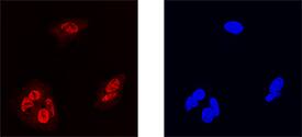
MafF in HepG2 Human Cell Line.
MafF was detected in immersion fixed HepG2 human hepatocellular carcinoma cell line using Goat Anti-Human MafF Antigen Affinity-purified Polyclonal Antibody (Catalog # AF3917) at 15 µg/mL for 3 hours at room temperature. Cells were stained using the NorthernLights™ 557-conjugated Anti-Goat IgG Secondary Antibody (left panel, red; Catalog # NL001) and counterstained with DAPI (right panel, blue). Specific staining was localized to nuclei. View our protocol for Fluorescent ICC Staining of Cells on Coverslips.Applications for Human MafF Antibody
Application
Recommended Usage
Immunocytochemistry
5-15 µg/mL
Sample: Immersion fixed HepG2 human hepatocellular carcinoma cell line
Sample: Immersion fixed HepG2 human hepatocellular carcinoma cell line
Western Blot
2 µg/mL
Sample: MDA-MB-468 human breast cancer cell line and SK-Mel-28 human malignant melanoma cell line
Sample: MDA-MB-468 human breast cancer cell line and SK-Mel-28 human malignant melanoma cell line
Formulation, Preparation, and Storage
Purification
Antigen Affinity-purified
Reconstitution
Reconstitute at 0.2 mg/mL in sterile PBS. For liquid material, refer to CoA for concentration.
Formulation
Lyophilized from a 0.2 μm filtered solution in PBS with Trehalose. *Small pack size (SP) is supplied either lyophilized or as a 0.2 µm filtered solution in PBS.
Shipping
Lyophilized product is shipped at ambient temperature. Liquid small pack size (-SP) is shipped with polar packs. Upon receipt, store immediately at the temperature recommended below.
Stability & Storage
Use a manual defrost freezer and avoid repeated freeze-thaw cycles.
- 12 months from date of receipt, -20 to -70 °C as supplied.
- 1 month, 2 to 8 °C under sterile conditions after reconstitution.
- 6 months, -20 to -70 °C under sterile conditions after reconstitution.
Background: MafF
Maf family members form a unique subclass of basic-leucine zipper (bZIP) transcription factors. Maf proteins are subdivided into two groupings: large, including
c‑Maf, Nrl, MafA, and MafB; and small, including MafF, MafG, and MafK. Large Mafs contain an N-terminal acidic domain important for transcriptional activation that is lacking in small Maf family members. Small Maf/Nrf2 heterodimers have been implicated in the regulation of antioxidant response element-dependent genes.
Long Name
v-maf Musculoaponeurotic Fibrosarcoma Oncogene Homolog F
Alternate Names
hMafF, transcription factor MafF, U-Maf, v-maf avian musculoaponeurotic fibrosarcoma oncogene family protein F, v-maf musculoaponeurotic fibrosarcoma (avian) oncogene family, protein F, V-maf musculoaponeurotic fibrosarcoma oncogene homolog F, v-maf musculoaponeurotic fibrosarcoma oncogene homolog F (avian)
Gene Symbol
MAFF
UniProt
Additional MafF Products
Product Documents for Human MafF Antibody
Product Specific Notices for Human MafF Antibody
For research use only
Loading...
Loading...
Loading...
Loading...
Loading...