
Human Integrin alpha8 Antibody
R&D Systems, part of Bio-Techne | Catalog # MAB6194

Scientific images may be resized, cropped, or adjusted for brightness or contrast for presentation purposes. These updates are limited to presentation and do not affect the underlying scientific interpretation of the data. Where available, additional source data may be provided upon request.
Conjugate
Catalog #
Key Product Details
Species Reactivity
Validated:
Human
Cited:
Human
Applications
Validated:
Flow Cytometry, Immunocytochemistry, CyTOF-ready
Cited:
Western Blot, Flow Cytometry, Bioassay
Label
Unconjugated
Antibody Source
Monoclonal Mouse IgG2B Clone # 481709
Product Specifications
Immunogen
E. coli-derived recombinant human Integrin alpha8
Glu610-Leu800
Accession # P53708
Glu610-Leu800
Accession # P53708
Specificity
Detects human Integrin alpha8 in direct ELISAs.
Clonality
Monoclonal
Host
Mouse
Isotype
IgG2B
Scientific Data Images for Human Integrin alpha8 Antibody
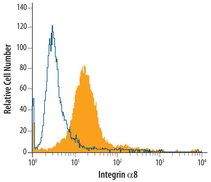
Detection of Integrin alpha8 in H4 Human Cell Line by Flow Cytometry.
H4 human neuroglioma cell line was stained with Mouse Anti-Human Integrin a8 Monoclonal Antibody (Catalog # MAB6194, filled histogram) or isotype control antibody (Catalog # MAB0041, open histogram), followed by Phycoerythrin-conjugated Anti-Mouse IgG Secondary Antibody (Catalog # F0102B).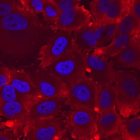
Integrin alpha8 in H4 Human Cell Line.
Integrin a8 was detected in immersion fixed H4 human neuroglioma cell line using Mouse Anti-Human Integrin a8 Monoclonal Antibody (Catalog # MAB6194) at 10 µg/mL for 3 hours at room temperature. Cells were stained using the NorthernLights™ 557-conjugated Anti-Mouse IgG Secondary Antibody (red; Catalog # NL007) and counterstained with DAPI (blue). Specific staining was localized to cell surfaces and cytoplasm. View our protocol for Fluorescent ICC Staining of Cells on Coverslips.
Detection of Integrin alpha 8 by Immunocytochemistry/ Immunofluorescence
Integrin-based subcellular adhesion structures on RGD and LDVP surfaces.Fab (100 nM for HP1/7 and 20 nM for the others) and Sir-actin (10 nM) were added 10 min before live-cell imaging of cells seeded for 1 h. a Integrin alphaV (AF488-13C2 Fab, green in the merged images) and actin stress fibers (SiR-actin, red in the merged images) on RGD-54pN. Intensity profiles (bottom right panel) are along the dashed line in the third enlarged image. Arbitrary units (a.u.). b Integrin alpha4 (AF488-HP1/7 Fab) and actin stress fibers (SiR-actin) on LDVP-54pN. c Integrin alpha4 (Cy3-HP1/7 Fab) and alphaV (AF647-13C2 Fab). d Paxillin (AF488, green) immunostaining of fixed cells imaged with SiR-actin (red) and alphaV (Cy3-13C2 Fab) or alpha4 (Cy3-HP1/7 Fab). e Extended open beta1 (Cy3-12G10 Fab) and closed beta1 (AF647-mAb13 Fab) on RGD-54pN. f Extended open beta1 (Cy3-12G10 Fab) and closed beta1 (AF647-mAb13 Fab) on LDVP-54pN. Boxed regions are magnified in the bottom row or right column. Scale bars: 10 µm (white), 2 µm (yellow). Source data are provided as a Source Data file. Image collected and cropped by CiteAb from the following open publication (https://pubmed.ncbi.nlm.nih.gov/36463259), licensed under a CC-BY license. Not internally tested by R&D Systems.Applications for Human Integrin alpha8 Antibody
Application
Recommended Usage
CyTOF-ready
Ready to be labeled using established conjugation methods. No BSA or other carrier proteins that could interfere with conjugation.
Flow Cytometry
2.5 µg/106 cells
Sample: H4 human neuroglioma cell line
Sample: H4 human neuroglioma cell line
Immunocytochemistry
8-25 µg/mL
Sample: Immersion fixed H4 human neuroglioma cell line
Sample: Immersion fixed H4 human neuroglioma cell line
Formulation, Preparation, and Storage
Purification
Protein A or G purified from hybridoma culture supernatant
Reconstitution
Sterile PBS to a final concentration of 0.5 mg/mL. For liquid material, refer to CoA for concentration.
Formulation
Lyophilized from a 0.2 μm filtered solution in PBS with Trehalose. *Small pack size (SP) is supplied either lyophilized or as a 0.2 µm filtered solution in PBS.
Shipping
Lyophilized product is shipped at ambient temperature. Liquid small pack size (-SP) is shipped with polar packs. Upon receipt, store immediately at the temperature recommended below.
Stability & Storage
Use a manual defrost freezer and avoid repeated freeze-thaw cycles.
- 12 months from date of receipt, -20 to -70 °C as supplied.
- 1 month, 2 to 8 °C under sterile conditions after reconstitution.
- 6 months, -20 to -70 °C under sterile conditions after reconstitution.
Background: Integrin alpha 8
Alternate Names
ITGA8
Gene Symbol
ITGA8
UniProt
Additional Integrin alpha 8 Products
Product Documents for Human Integrin alpha8 Antibody
Product Specific Notices for Human Integrin alpha8 Antibody
For research use only
Loading...
Loading...
Loading...
Loading...
Loading...
Loading...