Human HIF-1 alpha/HIF1A Antibody
R&D Systems, part of Bio-Techne | Catalog # MAB19352


Key Product Details
Validated by
Species Reactivity
Applications
Label
Antibody Source
Product Specifications
Immunogen
Arg575-Asn826
Accession # Q16665.1
Specificity
Clonality
Host
Isotype
Scientific Data Images for Human HIF-1 alpha/HIF1A Antibody
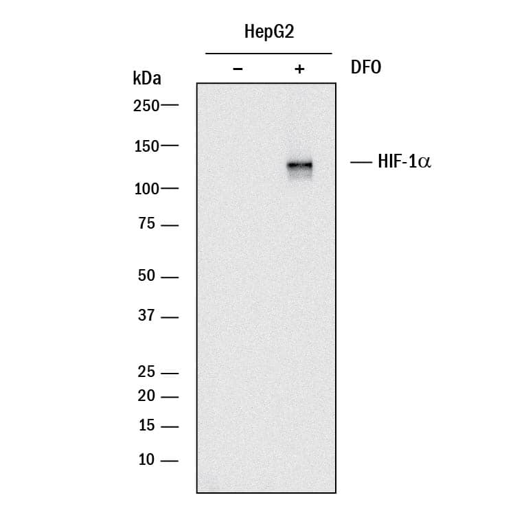
Detection of Human HIF-1 alpha/HIF1A by Western Blot.
Western blot shows lysates of HepG2 human hepatocellular carcinoma cell line untreated (-) or treated (+) with 1 mM DFO for overnight. PVDF membrane was probed with 2 µg/mL of Rabbit Anti-Human HIF-1 alpha/HIF1A Monoclonal Antibody (Catalog # MAB19352) followed by HRP-conjugated Anti-Rabbit IgG Secondary Antibody (Catalog # HAF008). A specific band was detected for HIF-1 alpha/HIF1A at approximately 120 kDa (as indicated). This experiment was conducted under reducing conditions and using Immunoblot Buffer Group 1.HIF-1 alpha/HIF1A in MCF-7 Human Cell Line.
HIF-1 alpha/HIF1A was detected in immersion fixed MCF-7 human breast cancer cell line using Rabbit Anti-Human HIF-1 alpha/HIF1A Monoclonal Antibody (Catalog # MAB19352) at 1.7 µg/mL for 3 hours at room temperature. Cells were stained using the NorthernLights™ 557-conjugated Anti-Rabbit IgG Secondary Antibody (red; Catalog # NL004) and counterstained with DAPI (blue). Specific staining was localized to nuclei. View our protocol for Fluorescent ICC Staining of Cells on Coverslips.HIF-1 alpha/HIF1A in HeLa Human Cell Line.
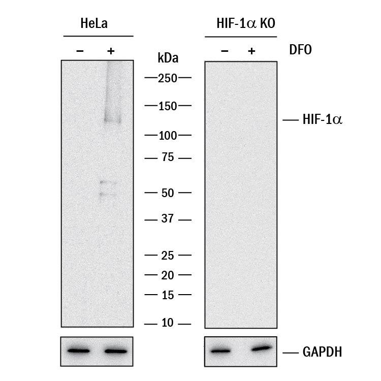
HIF-1 alpha/HIF1A was detected in immersion fixed HeLa human cervical epithelial carcinoma cell line treated with DFO (left panel, positive stain) or HeLa knockouts treated with DFO (right panel, negative stain) using Rabbit Anti-Human HIF-1 alpha/HIF1A Monoclonal Antibody (Catalog # MAB19352) at 1 µg/mL for 3 hours at room temperature. Cells were stained using a Goat anti-Rabbit IgG Secondary Antibody, Alexa Fluor 488 (green) and counterstained with DAPI (blue). Specific staining was localized to nuclei. View our protocol for Fluorescent ICC Staining of Cells on Coverslips.Applications for Human HIF-1 alpha/HIF1A Antibody
Immunocytochemistry
Sample: Immersion fixed MCF-7 human breast cancer cell line and HeLa human cervical epithelial carcinoma cell line treated with DFO and HeLa knockouts treated with DFO
Immunohistochemistry
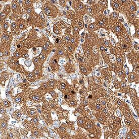
Sample: Immersion fixed paraffin-embedded sections of human liver
Knockout Validated
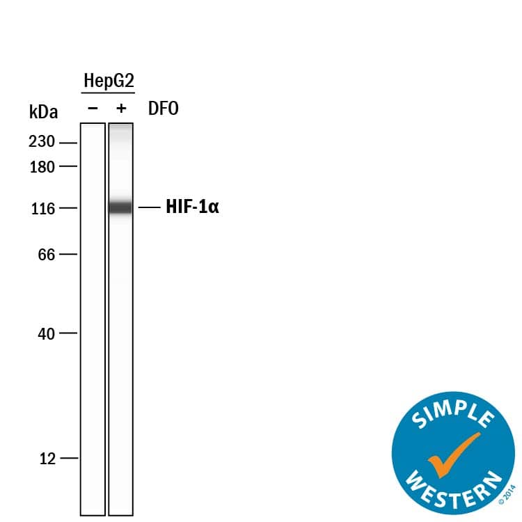
Simple Western
Sample: HepG2 human hepatocellular carcinoma cell line treated with DFO
Western Blot
Sample: HIF-1 alpha/HIF1A human hepatocellular carcinoma cell line treated with DFO
Formulation, Preparation, and Storage
Purification
Reconstitution
Formulation
Shipping
Stability & Storage
- 12 months from date of receipt, -20 to -70 °C as supplied.
- 1 month, 2 to 8 °C under sterile conditions after reconstitution.
- 6 months, -20 to -70 °C under sterile conditions after reconstitution.
Background: HIF-1 alpha/HIF1A
The hypoxia-inducible transcription factor 1 alpha (HIF-1 alpha) is the regulated member of the transcription factor heterodimer HIF-1. HIF-1 binds to hypoxia-response elements (HREs) in the promoters of many genes involved in adapting to an environment of insufficient oxygen or hypoxia. Hypoxic tissue environments occur in vascular and pulmonary diseases as well as cancer, which illustrates the broad impact of gene regulation by HIF-1 alpha.
Long Name
Alternate Names
Gene Symbol
UniProt
Additional HIF-1 alpha/HIF1A Products
Product Documents for Human HIF-1 alpha/HIF1A Antibody
Product Specific Notices for Human HIF-1 alpha/HIF1A Antibody
For research use only


