
Human Flt-3/Flk-2 APC-conjugated Antibody
R&D Systems, part of Bio-Techne | Catalog # FAB812A

Conjugate
Catalog #
Key Product Details
Species Reactivity
Human
Applications
Flow Cytometry
Label
Allophycocyanin (Excitation = 620-650 nm, Emission = 660-670 nm)
Antibody Source
Monoclonal Mouse IgG1 Clone # 66903
Product Specifications
Immunogen
Mouse myeloma cell line NS0-derived recombinant human Flt‑3/Flk‑2
Asn27-Asn541
Accession # AAA18947
Asn27-Asn541
Accession # AAA18947
Specificity
Detect human Flt‑3/Flk‑2 in direct ELISAs. In direct ELISAs, no cross-reactivity with recombinant mouse Flt-3, recombinant human (rh) SCF R, rhPDGF R alpha, or rhPDGF R beta is observed.
Clonality
Monoclonal
Host
Mouse
Isotype
IgG1
Scientific Data Images for Human Flt-3/Flk-2 APC-conjugated Antibody
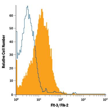
Detection of Flt‑3/Flk‑2 in THP‑1 Human Cell Line by Flow Cytometry.
THP-1 human acute monocytic leukemia cell line was stained with Mouse Anti-Human Flt-3/Flk-2 APC-conjugated Monoclonal Antibody (Catalog # FAB812A, filled histogram) or isotype control antibody (Catalog # IC002A, open histogram). View our protocol for Staining Membrane-associated Proteins.Applications for Human Flt-3/Flk-2 APC-conjugated Antibody
Application
Recommended Usage
Flow Cytometry
10 µL/106 cells
Sample: THP‑1 human acute monocytic leukemia cell line
Sample: THP‑1 human acute monocytic leukemia cell line
Spectra Viewer
Plan Your Experiments
Use our spectra viewer to interactively plan your experiments, assessing multiplexing options. View the excitation and emission spectra for our fluorescent dye range and other commonly used dyes.
Spectra Viewer
Formulation, Preparation, and Storage
Purification
Protein A or G purified from hybridoma culture supernatant
Formulation
Supplied in a saline solution containing BSA and Sodium Azide.
Shipping
The product is shipped with polar packs. Upon receipt, store it immediately at the temperature recommended below.
Stability & Storage
Protect from light. Do not freeze.
- 12 months from date of receipt, 2 to 8 °C as supplied.
Background: Flt-3/Flk-2
The Flt-3 (fms-Like Tyrosine kinase) Receptor, also known as Flk-2 (Fetal Liver Kinase) and Stk-1(Stem cell Tyrosine Kinase) is a member of the class III subfamily of receptor tyrosine kinases that also includes KIT, the receptor for SCF and FMS, the receptor for M-CSF. The extracellular region of these receptors contains five immunoglobulin-like domains and the intracellular region contains a split kinase domain. Human Flt-3 cDNA encodes a 993 amino acid (aa) residue type I membrane protein with a 26 aa residue signal peptide, a 515 aa extracellular domain with 10 potential N-linked glycosylation sites, a 21 aa residue transmembrane domain and a 431 aa residue cytoplasmic domain. Mouse Flt-3 has also been cloned and shown to share 85% amino acid sequence identity with human Flt-3. Flt-3 expression has been detected in various tissues, including placenta, gonads, and tissues of nervous and hematopoietic origin. Among hematopoietic cells, the expression of Flt-3 was found to be restricted to the highly enriched stem/progenitor cell populations. The ligand for Flt-3 (FL) has been identified to be a transmembrane protein with structural homology to M-CSF and SCF. Recombinant soluble Flt-3/Fc chimeric protein has been shown to bind FL with high affinity and is a potent FL antagonist.
References
- Rosnet, O. et al. (1996) Acta. Haemato. 95:218.
- Drexler, H.G. (1996) Leukemia 10:588.
Long Name
fms-like Tyrosine Kinase 3
Alternate Names
CD135, Flk-2, Flt3, STK-1
Gene Symbol
FLT3
UniProt
Additional Flt-3/Flk-2 Products
Product Documents for Human Flt-3/Flk-2 APC-conjugated Antibody
Product Specific Notices for Human Flt-3/Flk-2 APC-conjugated Antibody
For research use only
Loading...
Loading...
Loading...
Loading...
Loading...