
Human Cytokeratin 7 Antibody
R&D Systems, part of Bio-Techne | Catalog # MAB11626

Conjugate
Catalog #
Key Product Details
Species Reactivity
Human
Applications
Immunohistochemistry, Western Blot, Immunocytochemistry, COMET
Label
Unconjugated
Antibody Source
Monoclonal Mouse IgG2B Clone # 1090519
Product Specifications
Immunogen
Synthetic Peptide
Specificity
Detects a synthetic peptide specific for human Cytokeratin 7 around amino acid 300 in Direct ELISA.
Clonality
Monoclonal
Host
Mouse
Isotype
IgG2B
Scientific Data Images for Human Cytokeratin 7 Antibody
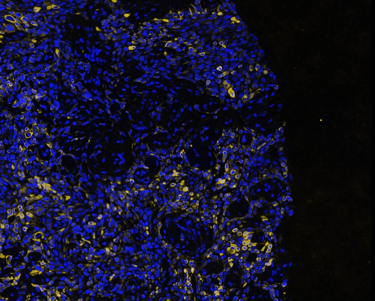
Detection of Cytokeratin 7 in human Pancreas Tumor via seqIF™ staining on COMET™
Cytokeratin 7 Peptide was detected in immersion fixed paraffin-embedded sections of human Pancreas Tumor using Mouse Anti-Human KRT7, Monoclonal Antibody (Catalog #MAB11626) at 25ug/mL at 37 ° Celsius for 4 minutes. Before incubation with the primary antibody, tissue underwent an all-in-one dewaxing and antigen retrieval preprocessing using PreTreatment Module (PT Module) and Dewax and HIER Buffer H (pH 9; Epredia Catalog # TA-999-DHBH). Tissue was stained using the Alexa Fluor™ 647 Goat anti-Mouse IgG Secondary Antibody at 1:200 at 37 ° Celsius for 2 minutes. (Yellow; Lunaphore Catalog # DR647MS) and counterstained with DAPI (blue; Lunaphore Catalog # DR100). Specific staining was localized to the cytoplasm. Protocol available in COMET™ Panel Builder.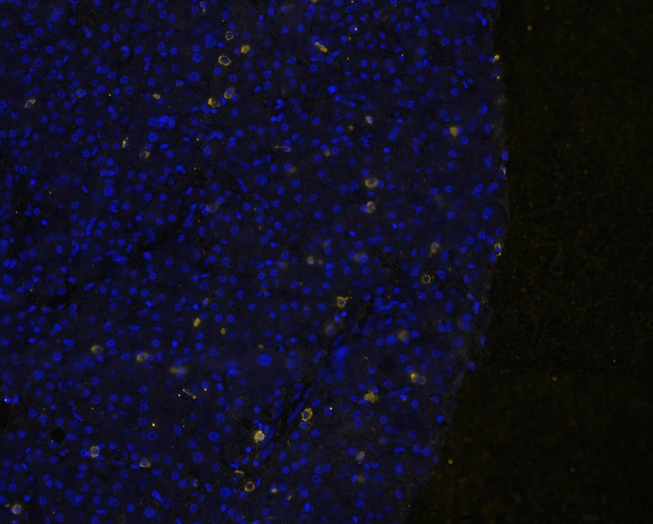
Detection of Cytokeratin 7 in human Liver Tumor via seqIF™ staining on COMET™
Cytokeratin 7 Peptide was detected in immersion fixed paraffin-embedded sections of human Liver Tumor using Mouse Anti-Human KRT7, Monoclonal Antibody (Catalog #MAB11626) at 25ug/mL at 37 ° Celsius for 4 minutes. Before incubation with the primary antibody, tissue underwent an all-in-one dewaxing and antigen retrieval preprocessing using PreTreatment Module (PT Module) and Dewax and HIER Buffer H (pH 9; Epredia Catalog # TA-999-DHBH). Tissue was stained using the Alexa Fluor™ 647 Goat anti-Mouse IgG Secondary Antibody at 1:200 at 37 ° Celsius for 2 minutes. (Yellow; Lunaphore Catalog # DR647MS) and counterstained with DAPI (blue; Lunaphore Catalog # DR100). Specific staining was localized to the cytoplasm. Protocol available in COMET™ Panel Builder.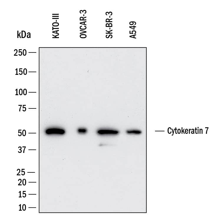
Detection of Human Cytokeratin 7 by Western Blot.
Western Blot shows lysates of KATO-III human gastric carcinoma cell line, OVCAR-3 human ovarian carcinoma cell line, SK-BR-3 human breast cancer cell line and A549 human lung carcinoma cell line. PVDF membrane was probed with 0.5 µg/ml of Mouse Anti-Human Cytokeratin 7 Monoclonal Antibody (Catalog # MAB11626) followed by HRP-conjugated Anti-Mouse IgG Secondary Antibody (Catalog # HAF018). A specific band was detected for Cytokeratin 7 at approximately 52 kDa (as indicated). This experiment was conducted under reducing conditions and using Western Blot Buffer Group 1.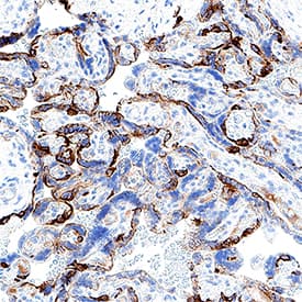
Applications for Human Cytokeratin 7 Antibody
Application
Recommended Usage
COMET
25 µg/mL
Sample: Immersion fixed paraffin-embedded sections of human liver tumor and pancreas tumor
Sample: Immersion fixed paraffin-embedded sections of human liver tumor and pancreas tumor
Immunocytochemistry
3-25 µg/mL
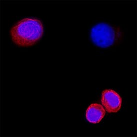
Sample: Immersion fixed SK-BR-3 human breast cancer cell line
Sample: Immersion fixed SK-BR-3 human breast cancer cell line
Immunohistochemistry
3-25 µg/mL
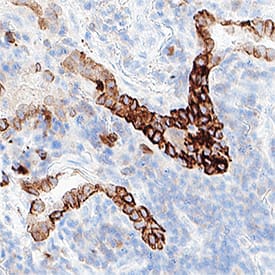
Sample: Immersion fixed paraffin-embedded sections of human lung cancer and human placenta
Sample: Immersion fixed paraffin-embedded sections of human lung cancer and human placenta
Western Blot
0.5 µg/mL
Sample: KATO-III human gastric carcinoma cell line, OVCAR-3 human ovarian carcinoma cell line, SK-BR-3 human breast cancer cell line and A549 human lung carcinoma cell line
Sample: KATO-III human gastric carcinoma cell line, OVCAR-3 human ovarian carcinoma cell line, SK-BR-3 human breast cancer cell line and A549 human lung carcinoma cell line
Formulation, Preparation, and Storage
Purification
Protein A or G purified from hybridoma culture supernatant
Reconstitution
Reconstitute lyophilized material at 0.2 mg/ml in sterile PBS. For liquid material, refer to CoA for concentration.
Formulation
Lyophilized from a 0.2 μm filtered solution in PBS with Trehalose.
Shipping
Lyophilized product is shipped at ambient temperature. Liquid small pack size (-SP) is shipped with polar packs. Upon receipt, store immediately at the temperature recommended below.
Stability & Storage
Use a manual defrost freezer and avoid repeated freeze-thaw cycles.
- 12 months from date of receipt, -20 to -70 °C as supplied.
- 1 month, 2 to 8 °C under sterile conditions after reconstitution.
- 6 months, -20 to -70 °C under sterile conditions after reconstitution.
Background: Cytokeratin 7
References
- Schweizer J, Bowden PE, Coulombe PA, Langbein L, Lane EB, Magin TM, Maltais L, Omary MB, Parry DA, Rogers MA, Wright MW. New consensus nomenclature for mammalian keratins. J Cell Biol. 2006 Jul 17;174(2):169-74. doi: 10.1083/jcb.200603161. Epub 2006 Jul 10. PMID: 16831889; PMCID: PMC2064177.
- Dum D, Menz A, Völkel C, De Wispelaere N, Hinsch A, Gorbokon N, Lennartz M, Luebke AM, Hube-Magg C, Kluth M, Fraune C, Möller K, Bernreuther C, Lebok P, Clauditz TS, Jacobsen F, Sauter G, Uhlig R, Wilczak W, Steurer S, Minner S, Marx AH, Simon R, Burandt E, Krech T. Cytokeratin 7 and cytokeratin 20 expression in cancer: A tissue microarray study on 15,424 cancers. Exp Mol Pathol. 2022 Jun;126:104762. doi: 10.1016/j.yexmp.2022.104762. Epub 2022 Apr 4. PMID: 35390310.
Alternate Names
CK-7, CK7, K2C7, K7
Gene Symbol
KRT7
Additional Cytokeratin 7 Products
Product Documents for Human Cytokeratin 7 Antibody
Product Specific Notices for Human Cytokeratin 7 Antibody
For research use only
Loading...
Loading...
Loading...
Loading...
Loading...