
Human Caspase-8 Antibody
R&D Systems, part of Bio-Techne | Catalog # MAB704

Scientific images may be resized, cropped, or adjusted for brightness or contrast for presentation purposes. These updates are limited to presentation and do not affect the underlying scientific interpretation of the data. Where available, additional source data may be provided upon request.
Conjugate
Catalog #
Key Product Details
Validated by
Biological Validation
Species Reactivity
Validated:
Human
Cited:
Human
Applications
Validated:
Western Blot
Cited:
Western Blot, Immunocytochemistry
Label
Unconjugated
Antibody Source
Monoclonal Mouse IgG2A Clone # 84131
Product Specifications
Immunogen
E. coli-derived recombinant human Caspase-8
aa 234-391
aa 234-391
Specificity
Detects human Caspase-8 precursor in Western blots and a 42 kDa doublet generated during apoptosis.
Clonality
Monoclonal
Host
Mouse
Isotype
IgG2A
Scientific Data Images for Human Caspase-8 Antibody
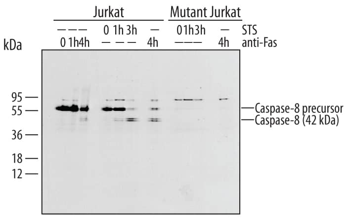
Detection of Human Caspase-8 by Western Blot.
Western blot shows lysates of Jurkat human acute T cell leukemia cell line untreated (-) or treated (+) with Human FAS Antigen Affinity-purified Polyclonal Antibody (Catalog # AF126) for indicated times. PVDF membrane was probed with 1 µg/mL of Human Caspase-8 Monoclonal Antibody (Catalog # MAB704), followed by HRP-conjugated Anti-Mouse IgG Secondary Antibody (Catalog # HAF007). Specific bands were detected for Caspase-8 at approximately 55 and 42 kDa (as indicated). This experiment was conducted under reducing conditions and using Immunoblot Buffer Group 4.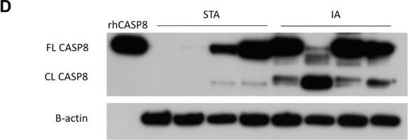
Detection of Caspase-8 by Western Blot
Validation of candidate diagnostic biomarkers in IA and STA samples. (A–C) Quantitative real-time PCR analysis of CASP8, PYCARD, and NOD2 mRNA levels in STA and IA tissue samples. All three genes were significantly upregulated in IA tissues compared with STA. (D) Western blot analysis of CASP8 protein expression. FL CASP8: full-length caspase-8; CL CASP8: cleaved caspase-8. Recombinant human CASP8 protein (rhCASP8) was used as a positive control. B-actin was used as a loading control. Data are shown as mean ± SD; ****p < 0.0001, ***p < 0.001. Image collected and cropped by CiteAb from the following open publication (https://pubmed.ncbi.nlm.nih.gov/42110963), licensed under a CC-BY license. Not internally tested by R&D Systems.Applications for Human Caspase-8 Antibody
Application
Recommended Usage
Western Blot
1 µg/mL
Sample: Anti Fas-treated Jurkat human acute T cell leukemia cell line
Sample: Anti Fas-treated Jurkat human acute T cell leukemia cell line
Reviewed Applications
Read 2 reviews rated 4.5 using MAB704 in the following applications:
Formulation, Preparation, and Storage
Purification
Protein A or G purified from ascites
Reconstitution
Reconstitute at 0.5 mg/mL in sterile PBS. For liquid material, refer to CoA for concentration.
Formulation
Lyophilized from a 0.2 μm filtered solution in PBS with Trehalose. *Small pack size (SP) is supplied either lyophilized or as a 0.2 µm filtered solution in PBS.
Shipping
Lyophilized product is shipped at ambient temperature. Liquid small pack size (-SP) is shipped with polar packs. Upon receipt, store immediately at the temperature recommended below.
Stability & Storage
Use a manual defrost freezer and avoid repeated freeze-thaw cycles.
- 12 months from date of receipt, -20 to -70 °C as supplied.
- 1 month, 2 to 8 °C under sterile conditions after reconstitution.
- 6 months, -20 to -70 °C under sterile conditions after reconstitution.
Background: Caspase-8
Caspase-8 (Cysteine-aspartic acid protease 8/Casp8; also MCH5 and FLICA) is a 28 kDa member of the peptidase C14A family of enzymes. It is known as the initiating caspase for the apoptotic cascade. Caspase-8 acts on procaspases-3, 4, 6, 7, 9 and 10, in addition to c-FLIPL and procaspase-8 itself. Human procaspase-8 alpha4 is a 58 kDa (predicted), 496 amino acid (aa) protein and contains two N-terminal death domains plus a catalytic site that utilizes His334Gly335 plus Cys377. Following death domain-containing receptor activation, proteolysis generates a 28 kDa heterodimer. This includes a p18 subunit (aa 234-391; or 217-374 of the 8a/standard form) noncovalently linked to a p10 subunit (aa 402-496; or 385-479 of the 8a/standard form). Association with another p18/p10 heterodimer generates active caspase-8. There are two common procaspase-8 isoform variants. One 53 kDa isoform (8b) shows a deletion of aa 103-134, while a second 55 kDa (8a) isoform shows a deletion of aa 103-134 with a 15 aa insertion after Lys183. The p18 and p10 subunits are 68% and 82% aa identical, human to mouse, respectively.
Additional Caspase-8 Products
Product Documents for Human Caspase-8 Antibody
Product Specific Notices for Human Caspase-8 Antibody
For research use only
Loading...
Loading...
Loading...
Loading...
Loading...