
Human alpha-Smooth Muscle Actin APC-conjugated Antibody
R&D Systems, part of Bio-Techne | Catalog # IC1420A

Conjugate
Catalog #
Key Product Details
Species Reactivity
Validated:
Human
Cited:
Human, Mouse
Applications
Validated:
Intracellular Staining by Flow Cytometry
Cited:
Immunohistochemistry, Flow Cytometry, Immunocytochemistry, FACS
Label
Allophycocyanin (Excitation = 620-650 nm, Emission = 660-670 nm)
Antibody Source
Monoclonal Mouse IgG2A Clone # 1A4
Product Specifications
Immunogen
alpha-Smooth Muscle Actin N-terminal Synthetic Decapeptide
Specificity
Detects human alpha-Smooth Muscle Actin.
Clonality
Monoclonal
Host
Mouse
Isotype
IgG2A
Scientific Data Images for Human alpha-Smooth Muscle Actin APC-conjugated Antibody
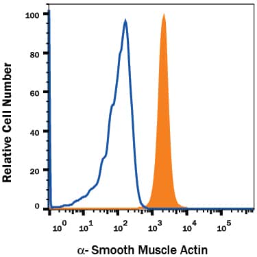
Detection of alpha‑Smooth Muscle Actin in Human Blood Monocytes by Flow Cytometry.
Human peripheral blood monocytes were stained with Mouse Anti-Human a-Smooth Muscle Actin APC-conjugated Monoclonal Antibody (Catalog # IC1420A, filled histogram) or isotype control antibody (Catalog # IC003A, open histogram). To facilitate intracellular staining, cells were fixed with Flow Cytometry Fixation Buffer (Catalog # FC004) and permeabilized with Flow Cytometry Permeabilization/Wash Buffer I (Catalog # FC005). View our protocol for Staining Intracellular Molecules.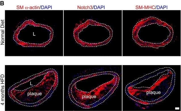
Detection of Mouse alpha-Smooth Muscle Actin by Immunohistochemistry
FGFR1 and TGF beta signaling activity in smooth muscle cells in a mouse atherosclerosis modelADissected mouse aorta demonstrating lipid‐rich plaques in brachiocephalic artery after 4 months of high‐fat diet compared to the normal diet in Apoe−/− mice. Panels b, d are cross sections of brachiocephalic artery from a, c stained with Oil Red O. L: lumen. Scale bar: 4 mm. n = 3 mice per group.BHistological analysis of mouse normal artery or atherosclerotic plaque in brachiocephalic artery with anti‐SM alpha‐actin, anti‐Notch3, and anti‐SM‐MHC antibodies. Nuclei were counterstained with DAPI (blue). L: lumen. Scale bar: 62 μm. n = 3 mice per group.C–FAnalysis of brachiocephalic artery of Apoe−/− mice maintained for 4 months on either normal or high‐fat diet using anti‐CD31 (green), anti‐p‐FGFR1 (red; C), anti‐FGFR1 (red; D), anti‐p‐Smad2 (red; E), and anti‐p‐Smad3 (red; F) antibodies. Nuclei were counterstained with DAPI (blue). Scale bar: 62 μm. L: lumen. M: media. n = 6 mice per group.G–JQuantification of the number of medial smooth muscle cells expressing p‐FGFR1, FGFR1, p‐Smad2, and p‐Smad3. ND: normal diet. HFD: high‐fat diet.Data information: All data shown as means ± SD (***P < 0.001 compared to ND, NS: not significant compared to ND; unpaired two‐tailed Student's t‐test). A full table of P‐values for this figure is shown in Appendix Table S1. Image collected and cropped by CiteAb from the following open publication (https://pubmed.ncbi.nlm.nih.gov/27189169), licensed under a CC-BY license. Not internally tested by R&D Systems.Applications for Human alpha-Smooth Muscle Actin APC-conjugated Antibody
Application
Recommended Usage
Intracellular Staining by Flow Cytometry
10 µL/106 cells
Sample: Human peripheral blood monocytes fixed with Flow Cytometry Fixation Buffer (Catalog # FC004) and permeabilized with Flow Cytometry Permeabilization/Wash Buffer I (Catalog # FC005)
Sample: Human peripheral blood monocytes fixed with Flow Cytometry Fixation Buffer (Catalog # FC004) and permeabilized with Flow Cytometry Permeabilization/Wash Buffer I (Catalog # FC005)
Spectra Viewer
Plan Your Experiments
Use our spectra viewer to interactively plan your experiments, assessing multiplexing options. View the excitation and emission spectra for our fluorescent dye range and other commonly used dyes.
Spectra Viewer
Formulation, Preparation, and Storage
Purification
Protein A or G purified from hybridoma culture supernatant
Formulation
Supplied in a saline solution containing BSA and Sodium Azide.
Shipping
The product is shipped with polar packs. Upon receipt, store it immediately at the temperature recommended below.
Stability & Storage
Protect from light. Do not freeze.
- 12 months from date of receipt, 2 to 8 °C as supplied.
Background: alpha-Smooth Muscle Actin
alpha‑Smooth Muscle Actin has been frequently used as a marker of smooth muscle differentiation (1, 2).
References
- Skalli, O. et al. (1986) J. Cell Biol. 103:2787.
- Oishi, K. et al. (2002) J. Physiol. 540:139.
Long Name
Actin, Alpha 2, Smooth Muscle, Aorta
Alternate Names
AAT6, ACTA2, Actin alpha 2, ACTSA, ACTVS, alphaSmooth Muscle Actin, MYMY5, SMA
Gene Symbol
ACTA2
Additional alpha-Smooth Muscle Actin Products
Product Documents for Human alpha-Smooth Muscle Actin APC-conjugated Antibody
Product Specific Notices for Human alpha-Smooth Muscle Actin APC-conjugated Antibody
For research use only
Loading...
Loading...
Loading...
Loading...
Loading...