T Cell Activation Phenotype Flow Cytometry Panel
T cells are activated upon exposure to their cognate antigen, or external stimuli in vitro. Expression patterns of early and late activation makers can help deepen the understanding of the host immune response. Use this validated multicolor flow cytometry panel to characterize your T cells for activation markers like CD25, CD69 and CD38.
Flow Cytometry Panel for Immunophenotyping of Activation T Cell Subsets
| Marker | Clone | Activated T Cells | Catalog # |
| CD3 | UCHT1* | mFluor™ Violet 450 | FAB100MFV450 |
| CD4 | 11830 | mFluor™ Violet 500 | FAB3791MFV500 |
| CD8 | 37006 | Alexa Fluor® 700 | FAB1509N |
| Live/Dead | (APC-Cy7) | ||
| CD25 | 24212 | mFluor™ Violet 610 | FAB1020MFV610 |
| CD69 | 298614 | PE | FAB23591P |
| CD38 | 240742 | Alexa Fluor® 700 | FAB2404R |
*Designate clones independently validated by HLDA.
Alexa Fluor® is registered trademark of Molecular Probes, Inc.
mFluor is a trademark of AAT Bioquest.
This multicolor flow cytometry panel was validated on human peripheral blood mononuclear cells (PBMCs).
Flow Cytometry Gating Strategy for Activated T Cell Panels
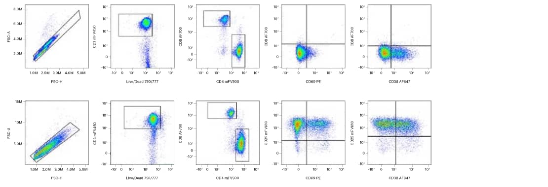
Multicolor flow cytometry panel to identify human Activation T cell subsets. PBMCs stimulated with ahCD3 (1 ug/mL; Cat# MAB11411-GMP) and ahCD28 (3 ug/mL; Cat# MAB11412-GMP) coupled to M-270 Dynabeads + IL-2 (200 U/mL; Cat# 202-GMP) for 14 days. Naïve T cells and Day 2 expanded T cells were stained with anti-human CD3 mFluor™ Violet 450, CD4 mFluor™ Violet 500, CD8 Alexa Fluor® 700, CD38 Alexa Fluor® 647, CD25 mFluor™ Violet 610, and CD69 PE. All antibodies are validated for flow cytometry. Gating strategy: Single Cells/Viable CD3+ cells/CD4+ vs. CD8+ cells. CD25, CD69, and CD38 expression were examined on Viable CD3+ cells assess T cell activation.
Staining Protocol For T Cell Activation Panel
Other supplies required
- PBS
- Flow Cytometry Staining Buffer (Catalog # FC001)
- Fc-block (blocking IgG)
- (Optional) Isotype Control Antibodies
- 5 mL Flow cytometry tubes
1. Wash human PBMCs (1 x 10⁶ cells per sample) with 2 mL of Staining Buffer (1X) (Catalog # FC001) or other BSA-containing buffer, by spinning at 300 x g for 5 minutes, using 5 mL flow cytometry tubes. Decant/aspirate supernatant.
2. Fc-block cells with blocking IgG (1 μg IgG/106 cells) for 10 minutes at room temperature.
3. Add previously titrated amount of each primary conjugated antibody. Vortex tubes.
| Marker | Fluorochrome | Volume/test (µL) |
| CD3 | mFluor™ Violet 450 | 5 |
| CD4 | mFluor™ Violet 500 | 5 |
| CD8 | Alexa Fluor® 700 | 5 |
| Live/Dead | (APC-Cy7) | 0.1 |
| CD25 | mFluor™ Violet 610 | 5 |
| CD69 | PE | 10 |
| CD38 | Alexa Fluor® 647 | 5 |
4. (Optional) To a separate tube, add 5 μL of each of the isotype control antibodies. Vortex tubes.
5. Incubate the mixtures for 30-45 minutes at room temperature in the dark.
6. At the end of the incubation, wash with 2 mL of Staining Buffer (1X), by spinning at 300 x g for 5 minutes. Decant/aspirate supernatant.
Additional Flow Cytometry Products and Resources
Products:
MagCellect™ Cell Selection Kits
Quality Control and Standardization Beads from Novus Biologicals
Resources:
Intracellular Staining with Alcohol Permeabilization Protocol
Intracellular Staining with Detergent Permeabilization Protocol
On-Demand Webinar: Demystifying Multi-parameter Flow Cytometry
On Demand Webinar: Turning Flow Cytometry Upside Down and Inside Out