
Mouse IgG2B Isotype Control Best Seller
R&D Systems, part of Bio-Techne | Catalog # MAB004


Scientific images may be resized, cropped, or adjusted for brightness or contrast for presentation purposes. These updates are limited to presentation and do not affect the underlying scientific interpretation of the data. Where available, additional source data may be provided upon request.
Key Product Details
Species Reactivity
Mouse
Applications
Control
Label
Unconjugated
Antibody Source
Monoclonal Mouse IgG2B Clone # 20116
Product Specifications
Immunogen
KLH
Specificity
Selected for use as mouse IgG2B isotype control. In direct ELISAs, does not cross-react with recombinant human (rh) 6Ckine, recombinant mouse (rm) 6Ckine, rhACT II (rhMIP-1 beta), rhBLC/BCA-1, rmBLC, rhBRAK, rmBRAK, rmC10, recombinant rat (rr) CINC-1, rrCINC-2 alpha, rrCINC-2 beta, rrCINC-3, rhCK beta8-1, rmCRG-2, rmCTACK, rhENA-78, rhEotaxin, rmEotaxin, rhEotaxin 2, rmEotaxin-2, rhEotaxin-3, rhFractalkine, rmFractalkine, rrFractalkine, rhGCP-2, rmGCP-2, rhGRO alpha, rhGRO beta, rhGRO gamma, rhHCC-1, rhHCC-4, rhI-309, rhIL-8, recombinant porcine IL-8, rhIP-10, rhI-TAC, rmI-TAC, rmJE, rmKC, rhLeukotactin-1, rrLIX, rhLymphotactin, rmLymphotactin, rmMARC, rhMCP-1, rhMCP-2, rhMCP-3, rhMCP-4, rmMCP-5, rhMDC, rmMDC, rhMIG, rmMIG, rhMIP-1 alpha, rmMIP-1 alpha, rmMIP-1 beta, rmMIP-1 gamma, rhMIP-1 delta, rmMIP-2, rhMIP-3 alpha, rmMIP-3 alpha, rrMIP-3 alpha, rhMIP-3 beta, rmMIP-3 beta, recombinant viral (rv) MIP-I, rvMIP-II, rvMIP-III, rhMPIF-1, rhNAP-2, rhParc, rhRANTES, rmRANTES, rhSDF-1 alpha, rmSDF-1 alpha, rhSDF-1 beta, rhTarc, rmTarc, rmTCA-3, rhTeck, rmTeck, rhVIC, or rmVIC.
Clonality
Monoclonal
Host
Mouse
Isotype
IgG2B
Scientific Data Images for Mouse IgG2B Isotype Control
Detection of Mouse IgG Control by Flow Cytometry
K562 human chronic myelogenous leukemia cell line was stained with Mouse Anti-Human ITFG1 Monoclonal Antibody (Catalog # MAB89001, filled histogram) or Mouse IgG Isotype Control Antibody (Catalog # MAB004, open histogram), followed by Allophycocyanin-conjugated Anti-Mouse IgG Secondary Antibody (Catalog F0101B).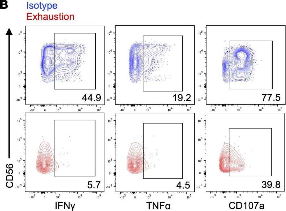
Detection of IgG2B by Flow Cytometry
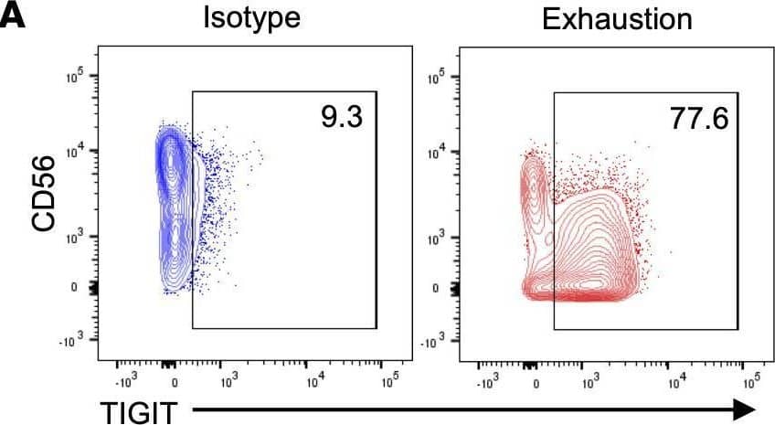
Prolonged stimulation through activating receptors induces NK cell exhaustion.(A) Schematic representing the in vitro model of exhaustion. Agonists of NKp46 (anti-NKp46) and NKG2D (MICA and MICB) were adsorbed onto tissue culture plates and used to stimulate NK cells for 7 days. Plate-bound isotype IgG served as a control. Both groups received 1 ng/mL IL-15. (B) NK cells harvested from isotype-coated and exhaustion plates (day 7) were incubated with K-562 targets for 4 hours (E/T: 2:1). Cytokine production (IFN-gamma and TNF-alpha) and degranulation (CD107a) were measured via flow cytometry. Isotype NK cells (top row) in blue, exhausted NK cells (bottom row) in red. E/T, effector/target. (C–E) Quantification of cytokine production and degranulation as percentage of parent population (live, CD3–CD56+ cells) and mean fluorescence intensity (MFI) (n = 4). Paired t tests were used for comparisons. **P < 0.01; ***P < 0.001; ****P < 0.0001. Image collected and cropped by CiteAb from the following open publication (https://insight.jci.org/articles/view/150079), licensed under a CC-BY license. Not internally tested by R&D Systems.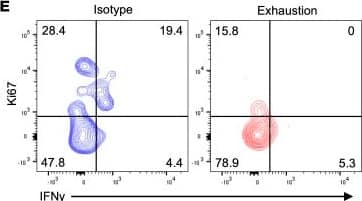
Detection of IgG2B by Flow Cytometry
Prolonged stimulation through activating receptors results in functional and proliferative defects in vivo.(A) Schematic representing experimental design: sublethally irradiated NSG mice were injected (i.v.) with HL-60-Luc2 cells and injected (i.v.) with NK cells 3 days later. NK cells had been incubated for 7 days on plates with either isotype IgG (control) or anti-NKp46 and MICA/B as previously described. (B) Fourteen days after NK cell injection, blood was drawn, and NK cells were counted via flow cytometry. NK cells were human CD45+CD56+CD3–. One-way ANOVA was used for comparisons (n = 8). *P ≤ 0.05. (C) HL-60 tumor burden was tracked via bioluminescent imaging (BLI), with day 21 data pictured. One-way ANOVA was used for comparisons (n = 8). ***P < 0.001. (D and E) Fourteen days after NK cell injection, NK cells were restimulated with K-562 leukemia cells for 4 hours as previously described. Ki67, IFN-gamma, TNF-alpha, and CD107a expression was assessed via flow cytometry (D). Paired t tests were used for comparisons (n = 8). *P ≤ 0.05. Representative data depict Ki67 and IFN-gamma expression following K-562 restimulation (E). Image collected and cropped by CiteAb from the following open publication (https://insight.jci.org/articles/view/150079), licensed under a CC-BY license. Not internally tested by R&D Systems.Applications for Mouse IgG2B Isotype Control
Application
Recommended Usage
Control
Negative control for use in conjunction with R&D Systems antibodies. Use at the same concentration as the detection antibody.
Reviewed Applications
Read 8 reviews rated 4.6 using MAB004 in the following applications:
Formulation, Preparation, and Storage
Purification
Protein A or G purified from hybridoma culture supernatant
Reconstitution
Reconstitute at 0.5 mg/mL in sterile PBS.
Formulation
Lyophilized from a 0.2 μm filtered solution in PBS with Trehalose.
Shipping
The product is shipped at ambient temperature. Upon receipt, store it immediately at the temperature recommended below.
Stability & Storage
Use a manual defrost freezer and avoid repeated freeze-thaw cycles.
- 12 months from date of receipt, -20 to -70 °C as supplied.
- 1 month, 2 to 8 °C under sterile conditions after reconstitution.
- 6 months, -20 to -70 °C under sterile conditions after reconstitution.
Background: IgG2B
R&D Systems offers a range of secondary antibodies and controls for flow cytometry, immunohistochemistry, and Western blotting. We provide species-specific secondary antibodies that are available with a variety of conjugated labels.
Our NorthernLights fluorescent secondary antibodies are bright and resistant to photobleaching. We are currently offering secondary antibodies recognizing mouse, rat, goat, sheep, and rabbit IgG as well as chicken IgY. These reagents are available with three distinct excitation and emission maxima, making them ideal for multi-color fluorescence microscopy.
Long Name
Immunoglobulin G2B Fc
Additional IgG2B Products
Product Documents for Mouse IgG2B Isotype Control
Product Specific Notices for Mouse IgG2B Isotype Control
For research use only
Loading...
Loading...
Loading...
Loading...