TRF-2 Antibody - BSA Free
Novus Biologicals, part of Bio-Techne | Catalog # NB110-57130
![Immunohistochemistry-Paraffin: TRF-2 Antibody - BSA Free [NB110-57130]](https://resources.bio-techne.com/images/products/TRF-2-Antibody-Immunohistochemistry-Paraffin-NB110-57130-img0007.jpg "Immunohistochemistry-Paraffin: TRF-2 Antibody - BSA Free [NB110-57130]")
![Immunocytochemistry/ Immunofluorescence: TRF-2 Antibody - BSA Free [NB110-57130]](https://resources.bio-techne.com/images/products/TRF-2-Antibody-Immunocytochemistry-Immunofluorescence-NB110-57130-img0011.jpg "Immunocytochemistry/ Immunofluorescence: TRF-2 Antibody - BSA Free [NB110-57130]")
Conjugate
Catalog #
Key Product Details
Validated by
Knockout/Knockdown
Species Reactivity
Validated:
Human, Mouse, Rat, Chinese Hamster, Primate
Cited:
Human, Mouse, Rat
Applications
Validated:
Immunohistochemistry, Immunohistochemistry-Paraffin, Western Blot, ELISA, Flow Cytometry, Flow (Intracellular), Immunocytochemistry/ Immunofluorescence, Simple Western, Immunoprecipitation, Chromatin Immunoprecipitation (ChIP), Dot Blot, Knockdown Validated
Cited:
Western Blot, ELISA, Immunocytochemistry/ Immunofluorescence, Immunoprecipitation, Chromatin Immunoprecipitation (ChIP), Chemotaxis, Cytometric Bead Assay Standard, FISH, Proximity Ligation Assay, IF/IHC, Knockdown
Label
Unconjugated
Antibody Source
Polyclonal Rabbit IgG
Format
BSA Free
Concentration
1.0 mg/ml
Product Specifications
Immunogen
This TRF-2 Antibody was developed against Baculovirus purified TRF2 protein.
Localization
Nuclear
Marker
Telomeres marker
Clonality
Polyclonal
Host
Rabbit
Isotype
IgG
Theoretical MW
59.6 kDa.
Disclaimer note: The observed molecular weight of the protein may vary from the listed predicted molecular weight due to post translational modifications, post translation cleavages, relative charges, and other experimental factors.
Disclaimer note: The observed molecular weight of the protein may vary from the listed predicted molecular weight due to post translational modifications, post translation cleavages, relative charges, and other experimental factors.
Scientific Data Images for TRF-2 Antibody - BSA Free
Immunocytochemistry/ Immunofluorescence: TRF-2 Antibody - BSA Free [NB110-57130]
Immunocytochemistry/Immunofluorescence: TRF-2 Antibody [NB110-57130] - HeLa cells were fixed for 10 minutes using 10% formalin and then permeabilized for 5 minutes using 1X TBS + 0.5% Triton X-100. The cells were incubated with antibody at a 1:200 dilution overnight at 4 degrees Celsius and detected with DyLight 488 (Green) at a 1:500 dilution. Alpha tubulin was used as a co-stain at a 1:1000 dilution and detected with Dylight 550 (Red). Nuclei were detected with DAPI (Blue) at 2.0 ug/ml in 1X PBS. Cells were imaged using a 40X objective.![Simple Western: TRF-2 AntibodyBSA Free [NB110-57130]](https://resources.bio-techne.com/images/products/TRF-2-Antibody-Simple-Western-NB110-57130-img0010.jpg "Simple Western: TRF-2 AntibodyBSA Free [NB110-57130]")
Simple Western: TRF-2 AntibodyBSA Free [NB110-57130]
Simple Western: TRF-2 Antibody [NB110-57130] - Lane view shows a specific band for TRF2 in 1.0 mg/mL of HeLa lysate. This experiment was performed under reducing conditions using the 12-230 kDa separation system.![Western Blot: TRF-2 AntibodyBSA Free [NB110-57130]](https://resources.bio-techne.com/images/products/TRF-2-Antibody-Western-Blot-NB110-57130-img0008.jpg "Western Blot: TRF-2 AntibodyBSA Free [NB110-57130]")
Western Blot: TRF-2 AntibodyBSA Free [NB110-57130]
Western Blot: TRF-2 Antibody [NB110-57130] - Analysis of HeLa whole cell lysate (A), HeLa nuclear cell lysate (B), k562 cell lysate (C), HepG2 cell lysate (D), NIH/3T3 cell lysate (E), CHO cell lysate (F), PC12 cell lysate (G), and Cos7 cell lysate (H) using antibody at a concentration of 2 ug/mL.![Immunohistochemistry-Paraffin: TRF-2 Antibody - BSA Free [NB110-57130]](https://resources.bio-techne.com/images/products/TRF-2-Antibody-Immunohistochemistry-Paraffin-NB110-57130-img0013.jpg "Immunohistochemistry-Paraffin: TRF-2 Antibody - BSA Free [NB110-57130]")
![Immunocytochemistry/ Immunofluorescence: TRF-2 Antibody - BSA Free [NB110-57130]](https://resources.bio-techne.com/images/products/TRF-2-Antibody-Immunocytochemistry-Immunofluorescence-NB110-57130-img0016.jpg "Immunocytochemistry/ Immunofluorescence: TRF-2 Antibody - BSA Free [NB110-57130]")
![Flow Cytometry: TRF-2 Antibody - BSA Free [NB110-57130]](https://resources.bio-techne.com/images/products/TRF-2-Antibody-Flow-Cytometry-NB110-57130-img0017.jpg "Flow Cytometry: TRF-2 Antibody - BSA Free [NB110-57130]")
![Immunocytochemistry/ Immunofluorescence: TRF-2 Antibody - BSA Free [NB110-57130]](https://resources.bio-techne.com/images/products/TRF-2-Antibody-Immunocytochemistry-Immunofluorescence-NB110-57130-img0015.jpg "Immunocytochemistry/ Immunofluorescence: TRF-2 Antibody - BSA Free [NB110-57130]")
![Immunohistochemistry-Paraffin: TRF-2 Antibody - BSA Free [NB110-57130]](https://resources.bio-techne.com/images/products/TRF-2-Antibody-Immunohistochemistry-Paraffin-NB110-57130-img0012.jpg "Immunohistochemistry-Paraffin: TRF-2 Antibody - BSA Free [NB110-57130]")
![Flow Cytometry: TRF-2 Antibody - BSA Free [NB110-57130]](https://resources.bio-techne.com/images/products/TRF-2-Antibody-Flow-Cytometry-NB110-57130-img0014.jpg "Flow Cytometry: TRF-2 Antibody - BSA Free [NB110-57130]")


Applications for TRF-2 Antibody - BSA Free
Application
Recommended Usage
Chromatin Immunoprecipitation (ChIP)
1:10-1:500
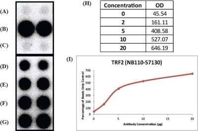
Dot Blot
reported in scientific literature (PMID 31026066)
ELISA
reported in scientific literature (PMID 31575660)
Flow Cytometry
1-5 ug/ml
Immunocytochemistry/ Immunofluorescence
1:50 - 1:200
Immunohistochemistry
1:200
Immunohistochemistry-Paraffin
1:200
Immunoprecipitation
1:10 - 1:500. Use reported in scientific literature
Knockdown Validated
reported in scientific literature (PMID 31026066)
Simple Western
1:25
Western Blot
1:2000 - 1:5000
Application Notes
In Western blot, a band at approx. 56 kDa is seen. In ICC/IF, nuclear staining was observed in HeLa cells. In IHC, nuclear staining was observed in xenografted human breast cancer tissue. Prior to immunostaining paraffin tissues, antigen retrieval with sodium citrate buffer (pH 6.0) is recommended.
In Simple Western only 10 - 15 uL of the recommended dilution is used per data point.
See Simple Western Antibody Database for Simple Western validation: Tested in HeLa lysate 1.0 mg/mL, separated by Size, antibody dilution of 1:25, apparent MW was 66 kDa. Separated by Size-Wes, Sally Sue/Peggy Sue.
The observed molecular weight of the protein may vary from the listed predicted molecular weight due to post translational modifications, post translation cleavages, relative charges, and other experimental factors.
Reviewed Applications
Read 2 reviews rated 5 using NB110-57130 in the following applications:
Formulation, Preparation, and Storage
Purification
Immunogen affinity purified
Formulation
PBS
Format
BSA Free
Preservative
0.02% Sodium Azide
Concentration
1.0 mg/ml
Shipping
The product is shipped with polar packs. Upon receipt, store it immediately at the temperature recommended below.
Stability & Storage
Store at 4C short term. Aliquot and store at -20C long term. Avoid freeze-thaw cycles.
Background: TRF-2
Both TRF2 and TRF1 bind to telomeric double stranded 5'-TTAGGG-3' DNA repeats, then recruit RAP1, TIN2, TPP1, and POT1 for the assembly of the shelterin complex. The telomeric association of TRF2 is greatly increased in the S phase of the cell cycle (2). Loss of TRF2 leads to telomere shortening, the DNA damage response, chromosomal instability, and replicative senescence. Interestingly, the contribution of TRF2 to telomere shortening via a telomerase-independent mechanism has also been reported (3). In conjunction with the exonuclease, Apollo, TRF2 protects telomeres during replication and negatively regulates the accumulation of DNA topoisomerase (TOP1, TOP2A and TOP2B).
TRF2 has been implicated in cancer, shown to be a major oncogene in telomerase-deficient mice. A link to Werner syndrome, a premature aging disease caused by the loss of WRN, has been reported based on TRF2 recruitment of WRN for processing of telomeric DNA (4). TRF2 expression is increased during human embryonic stem cell differentiation and has been shown to interact with Repressor Element-1 Silencing Transcription Factor (REST), protecting it from proteasomal degradation (5).
References
1. Grammatikakis, I., Zhang, P., Mattson, M. P., & Gorospe, M. (2016). The long and the short of TRF2 in neurogenesis. Cell cycle (Georgetown, Tex.), 15(22), 3026-3032. PMID: 27565210
2. Li, F., Kim, H., Ji, Z., Zhang, T., Chen, B., Ge, Y., Hu, Y., Feng, X., Han, X., Xu, H., Zhang, Y., Yu, H., Liu, D., Ma, W., & Songyang, Z. (2018). The BUB3-BUB1 Complex Promotes Telomere DNA Replication. Molecular cell, 70(3), 395-407. PMID: 29727616
3. Ancelin, K., Brunori, M., Bauwens, S., Koering, C. E., Brun, C., Ricoul, M., Pommier, J. P., Sabatier, L., & Gilson, E. (2002). Targeting assay to study the cis functions of human telomeric proteins: evidence for inhibition of telomerase by TRF1 and for activation of telomere degradation by TRF2. Molecular and cellular biology, 22(10), 3474-3487. PMID: 11971978
4. Machwe A, Xiao L, & Orren DK. (2004) TRF2 recruits the Werner syndrome (WRN) exonuclease for processing of telomeric DNA. Oncogene. 23(1):149-56. PMID: 14712220.
5. Diotti, R., & Loayza, D. (2011). Shelterin complex and associated factors at human telomeres. Nucleus (Austin, Tex.), 2(2), 119-135. PMID: 21738835
Long Name
Telomeric Repeat Binding Factor 2
Alternate Names
TERF2, TRBF2, TRF2
Gene Symbol
TERF2
Additional TRF-2 Products
Product Documents for TRF-2 Antibody - BSA Free
Product Specific Notices for TRF-2 Antibody - BSA Free
This product is for research use only and is not approved for use in humans or in clinical diagnosis. Primary Antibodies are guaranteed for 1 year from date of receipt.
Loading...
Loading...
Loading...
Loading...
Loading...
Loading...