
Human SSBP1 Antibody
R&D Systems, part of Bio-Techne | Catalog # AF6588


Scientific images may be resized, cropped, or adjusted for brightness or contrast for presentation purposes. These updates are limited to presentation and do not affect the underlying scientific interpretation of the data. Where available, additional source data may be provided upon request.
Conjugate
Catalog #
Key Product Details
Species Reactivity
Validated:
Human
Cited:
Human
Applications
Validated:
Western Blot, Immunocytochemistry
Cited:
Western Blot
Label
Unconjugated
Antibody Source
Polyclonal Sheep IgG
Product Specifications
Immunogen
E. coli-derived recombinant human SSBP1
Met1-Glu148
Accession # Q04837
Met1-Glu148
Accession # Q04837
Specificity
Detects human SSBP1 in direct ELISAs and Western blots.
Clonality
Polyclonal
Host
Sheep
Isotype
IgG
Scientific Data Images for Human SSBP1 Antibody
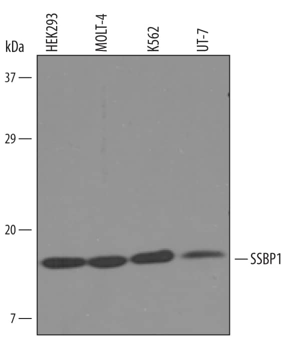
Detection of Human SSBP1 by Western Blot.
Western blot shows lysates of HEK293 human embryonic kidney cell line, MOLT-4 human acute lymphoblastic leukemia cell line, K562 human chronic myelogenous leukemia cell line, and UT-7 human acute myeloid leukemia cell line. PVDF Membrane was probed with 1 µg/mL of Sheep Anti-Human SSBP1 Antigen Affinity-purified Polyclonal Antibody (Catalog # AF6588) followed by HRP-conjugated Anti-Sheep IgG Secondary Antibody (Catalog # HAF016). A specific band was detected for SSBP1 at approximately 18 kDa (as indicated). This experiment was conducted under reducing conditions and using Immunoblot Buffer Group 8.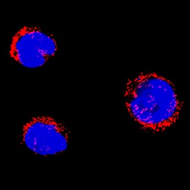
SSBP1 in HL‑60 Human Cell Line.
SSBP1 was detected in immersion fixed HL-60 human acute promyelocytic leukemia cell line using Sheep Anti-Human SSBP1 Antigen Affinity-purified Polyclonal Antibody (Catalog # AF6588) at 5 µg/mL for 3 hours at room temperature. Cells were stained using the NorthernLights™ 557-conjugated Anti-Sheep IgG Secondary Antibody (red; Catalog # NL010) and counterstained with DAPI (blue). Specific staining was localized to cytoplasm. View our protocol for Fluorescent ICC Staining of Non-adherent Cells.
Detection of SSBP1 by Western Blot
Outside–in degradation of mitochondrial proteins.A, mitochondrial proteins were enriched among significantly regulated proteins. B, abundance profiles of significantly regulated proteins from various mitochondrial subcompartments show trends dependent on parkin activity. Indicated are size of total annotated proteins (left; dark profiles) and those after filtering (right; bright profiles); four mitochondrial proteins are of unknown or dual sublocalization (data not shown). C, average profile of each cluster sorted by half maximum in combination with overrepresentation analysis of mitochondrial subcompartments allows for the discrimination of “early” degraded MOM, “intermediate” degraded MOM/IMS/MIM, and “late” degraded MIM/matrix proteins. D, Western blot detection of mitochondrial subcompartment proteins during mitophagy validates stepwise degradation of mitochondrial subcompartments. IMS, inter membrane space; Mito, mitochondrion; MIM, mitochondrial inner membrane; MOM, mitochondrial outer membrane. Image collected and cropped by CiteAb from the following open publication (https://linkinghub.elsevier.com/retrieve/pii/S1535947621001638), licensed under a CC-BY license. Not internally tested by R&D Systems.

Applications for Human SSBP1 Antibody
Application
Recommended Usage
Immunocytochemistry
5-15 µg/mL
Sample: Immersion fixed HL-60 human acute promyelocytic leukemia cell line
Sample: Immersion fixed HL-60 human acute promyelocytic leukemia cell line
Western Blot
1 µg/mL
Sample: HEK293 human embryonic kidney cell line, MOLT‑4 human acute lymphoblastic leukemia cell line, K562 human chronic myelogenous leukemia cell line, and UT‑7 human acute myeloid leukemia cell line
Sample: HEK293 human embryonic kidney cell line, MOLT‑4 human acute lymphoblastic leukemia cell line, K562 human chronic myelogenous leukemia cell line, and UT‑7 human acute myeloid leukemia cell line
Reviewed Applications
Read 1 review rated 4 using AF6588 in the following applications:
Formulation, Preparation, and Storage
Purification
Antigen Affinity-purified
Reconstitution
Sterile PBS to a final concentration of 0.2 mg/mL. For liquid material, refer to CoA for concentration.
Formulation
Lyophilized from a 0.2 μm filtered solution in PBS with Trehalose. *Small pack size (SP) is supplied either lyophilized or as a 0.2 µm filtered solution in PBS.
Shipping
Lyophilized product is shipped at ambient temperature. Liquid small pack size (-SP) is shipped with polar packs. Upon receipt, store immediately at the temperature recommended below.
Stability & Storage
Use a manual defrost freezer and avoid repeated freeze-thaw cycles.
- 12 months from date of receipt, -20 to -70 °C as supplied.
- 1 month, 2 to 8 °C under sterile conditions after reconstitution.
- 6 months, -20 to -70 °C under sterile conditions after reconstitution.
Background: SSBP1
Long Name
Single-stranded DNA Binding Protein Motochondrial
Alternate Names
mitochondrial, MtSSB, single-stranded DNA binding protein 1
Gene Symbol
SSBP1
UniProt
Additional SSBP1 Products
Product Documents for Human SSBP1 Antibody
Product Specific Notices for Human SSBP1 Antibody
For research use only
Loading...
Loading...
Loading...
Loading...
Loading...