
Human Phospho-STAT2 (Y690) Antibody
R&D Systems, part of Bio-Techne | Catalog # MAB2890
Recombinant Monoclonal Antibody.

Scientific images may be resized, cropped, or adjusted for brightness or contrast for presentation purposes. These updates are limited to presentation and do not affect the underlying scientific interpretation of the data. Where available, additional source data may be provided upon request.
Key Product Details
Validated by
Biological Validation
Species Reactivity
Validated:
Human
Cited:
Human
Applications
Validated:
Western Blot, Simple Western
Cited:
Western Blot
Label
Unconjugated
Antibody Source
Recombinant Monoclonal Rabbit IgG Clone # 1021D
Product Specifications
Immunogen
Phosphopeptide containing the human STAT2 Y690 site
Specificity
Detects human phospho-STAT2 (Y690) in direct ELISAs and Western blots.
Clonality
Monoclonal
Host
Rabbit
Isotype
IgG
Scientific Data Images for Human Phospho-STAT2 (Y690) Antibody
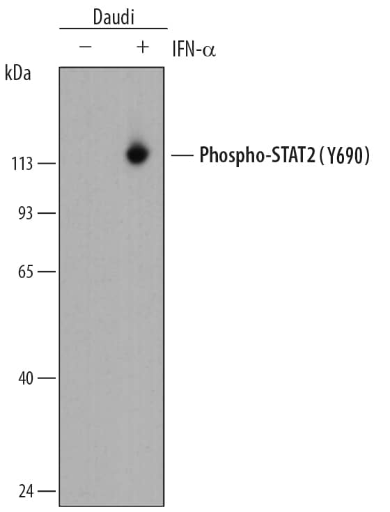
Detection of Human Phospho-STAT2 (Y690) by Western Blot.
Western blot shows lysates of Daudi human Burkitt's lymphoma cell line untreated (-) or treated (+) with 500 U/mL Recombinant Human IFN-aA (11100-1) for 20 minutes. PVDF membrane was probed with 0.05 µg/mL of Rabbit Anti-Human Phospho-STAT2 (Y690) Monoclonal Antibody (Catalog # MAB2890) followed by HRP-conjugated Anti-Rabbit IgG Secondary Antibody (HAF008). A specific band was detected for Phospho-STAT2 (Y690) at approximately 113 kDa (as indicated). This experiment was conducted under reducing conditions and using Immunoblot Buffer Group 1.
Detection of Human Phospho-STAT2 (Y690) by Simple WesternTM.
Simple Western lane view shows lysates of Daudi human Burkitt's lymphoma cell line untreated (-) or treated (+) with 500 U/mL Recombinant Human IFN-aA (11100-1) for 20 minutes, loaded at 0.2 mg/mL. A specific band was detected for Phospho-STAT2 (Y690) at approximately 111 kDa (as indicated) using 0.5 µg/mL of Rabbit Anti-Human Phospho-STAT2 (Y690) Monoclonal Antibody (Catalog # MAB2890). This experiment was conducted under reducing conditions and using the 12-230 kDa separation system.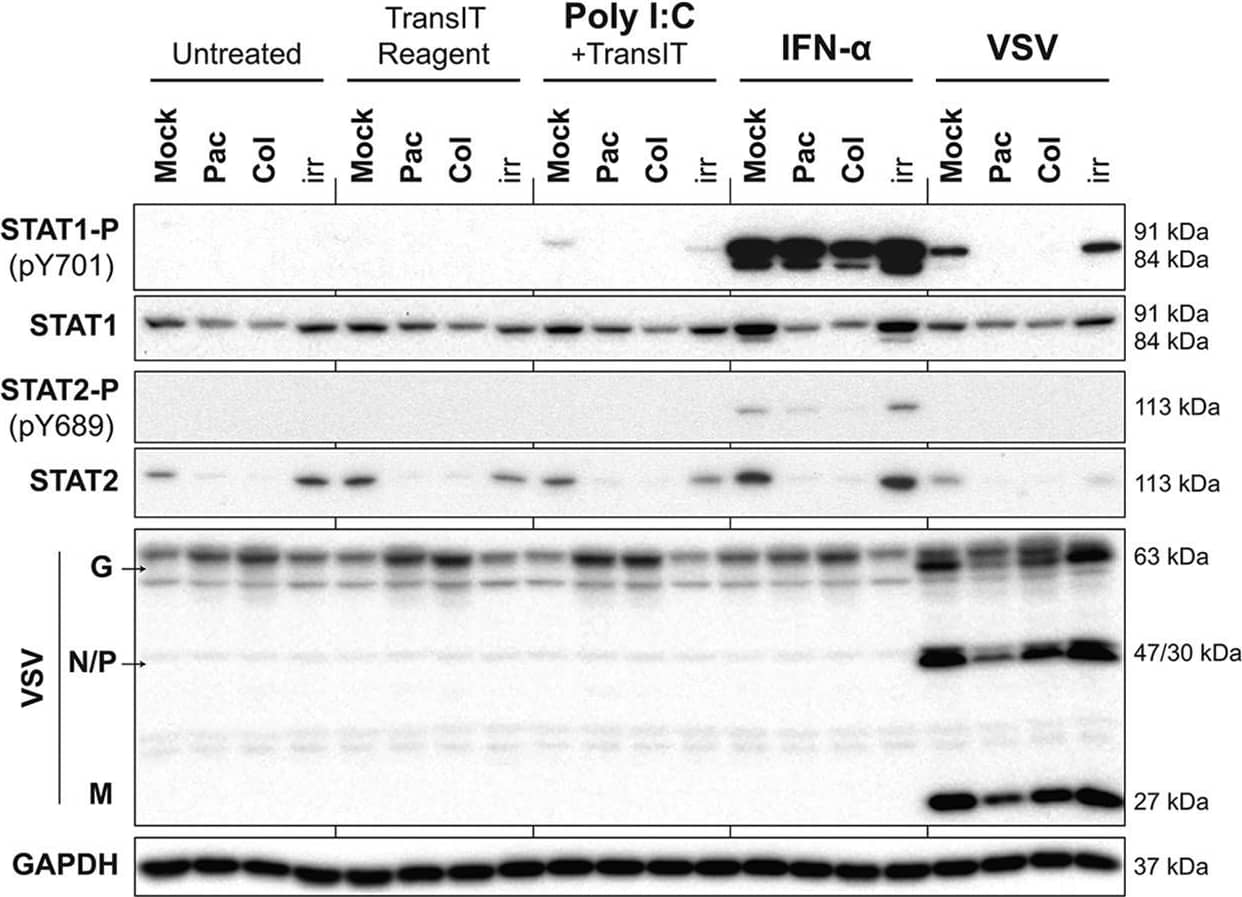
Detection of Human STAT2 by Western Blot
Induction of type I IFN signaling by viral and nonviral stimuli is inhibited in G2/M-arrested cells. Suit2 cells were treated for 25 h with the vehicle, paclitaxel, colchicine, or ruxolitinib at 500 nM. Cells were then treated with the vehicle (untreated), TransIT reagent (0.5%, vol/vol), poly(I:C) at 10 μg/ml plus TransIT reagent, IFN-alpha at 5,000 U/ml, or VSV at an MOI of 30 based on titration on BHK-21 cells. VSV was aspirated 1 h later, and medium was added to infected wells. Cells remained in treatment for a total of 4 h, after which total protein was isolated. Western blot results for STAT1 and -2 proteins and their phosphorylated forms are shown in addition to VSV proteins. GAPDH was used to confirm that protein loading was the same across the gel. Protein names and protein sizes in kilodaltons are indicated on the left and right, respectively. Image collected and cropped by CiteAb from the following publication (https://pubmed.ncbi.nlm.nih.gov/30487274), licensed under a CC-BY license. Not internally tested by R&D Systems.Applications for Human Phospho-STAT2 (Y690) Antibody
Application
Recommended Usage
Simple Western
0.5 µg/mL
Sample: Daudi human Burkitt's lymphoma cell line treated with Recombinant Human IFN‑ alphaA (Catalog # 11100-1)
Sample: Daudi human Burkitt's lymphoma cell line treated with Recombinant Human IFN‑ alphaA (Catalog # 11100-1)
Western Blot
0.05 µg/mL
Sample: Daudi human Burkitt's lymphoma cell line treated with Recombinant Human IFN‑ alphaA (Catalog # 11100-1)
Sample: Daudi human Burkitt's lymphoma cell line treated with Recombinant Human IFN‑ alphaA (Catalog # 11100-1)
Formulation, Preparation, and Storage
Purification
Protein A or G purified from cell culture supernatant
Reconstitution
Reconstitute at 0.5 mg/mL in sterile PBS. For liquid material, refer to CoA for concentration.
Formulation
Lyophilized from a 0.2 μm filtered solution in PBS with Trehalose. See Certificate of Analysis for details.
*Small pack size (-SP) is supplied either lyophilized or as a 0.2 µm filtered solution in PBS.
*Small pack size (-SP) is supplied either lyophilized or as a 0.2 µm filtered solution in PBS.
Shipping
Lyophilized product is shipped at ambient temperature. Liquid small pack size (-SP) is shipped with polar packs. Upon receipt, store immediately at the temperature recommended below.
Stability & Storage
Use a manual defrost freezer and avoid repeated freeze-thaw cycles.
- 12 months from date of receipt, -20 to -70 °C as supplied.
- 1 month, 2 to 8 °C under sterile conditions after reconstitution.
- 6 months, -20 to -70 °C under sterile conditions after reconstitution.
Background: STAT2
STAT2 (signal transducer and activator of transcription 2) is a 113 kDa member of the STAT family of cytoplasmic transcription factors. STAT members generally mediate cytokine, growth factor and hormone receptor signal transduction. STAT2 is associated with type I ( alpha- and beta-) interferon signaling. All STATs contain an N-terminal oligomerization domain, a DNA-binding domain, and an SH2-association region. STAT2 is phosphorylated at Y690 by receptor-associated Janus kinases (JAKs) leading to STAT2 dimerization and subsequent translocation to the nucleus to activate gene transcription.
Long Name
Signal Transducer and Activator of Transcription 2
Alternate Names
interferon alpha induced transcriptional activator, ISGF-3, MGC59816, P113, signal transducer and activator of transcription 2, signal transducer and activator of transcription 2, 113kD, signal transducer and activator of transcription 2, 113kDa, STAT113
Gene Symbol
STAT2
Additional STAT2 Products
Product Documents for Human Phospho-STAT2 (Y690) Antibody
Product Specific Notices for Human Phospho-STAT2 (Y690) Antibody
For research use only
Loading...
Loading...
Loading...
Loading...
Loading...
Loading...