
Human Phospho-ErbB3/Her3 (Y1262) Antibody
R&D Systems, part of Bio-Techne | Catalog # AF5817

Scientific images may be resized, cropped, or adjusted for brightness or contrast for presentation purposes. These updates are limited to presentation and do not affect the underlying scientific interpretation of the data. Where available, additional source data may be provided upon request.
Conjugate
Catalog #
Key Product Details
Validated by
Biological Validation
Species Reactivity
Validated:
Human
Cited:
Human, Mouse
Applications
Validated:
Western Blot
Cited:
Western Blot
Label
Unconjugated
Antibody Source
Polyclonal Rabbit IgG
Product Specifications
Immunogen
Phosphopeptide containing human ErbB3 Y1262 site
Specificity
Detects human ErbB3 when phosphorylated at Y1262 in Western blots.
Clonality
Polyclonal
Host
Rabbit
Isotype
IgG
Scientific Data Images for Human Phospho-ErbB3/Her3 (Y1262) Antibody
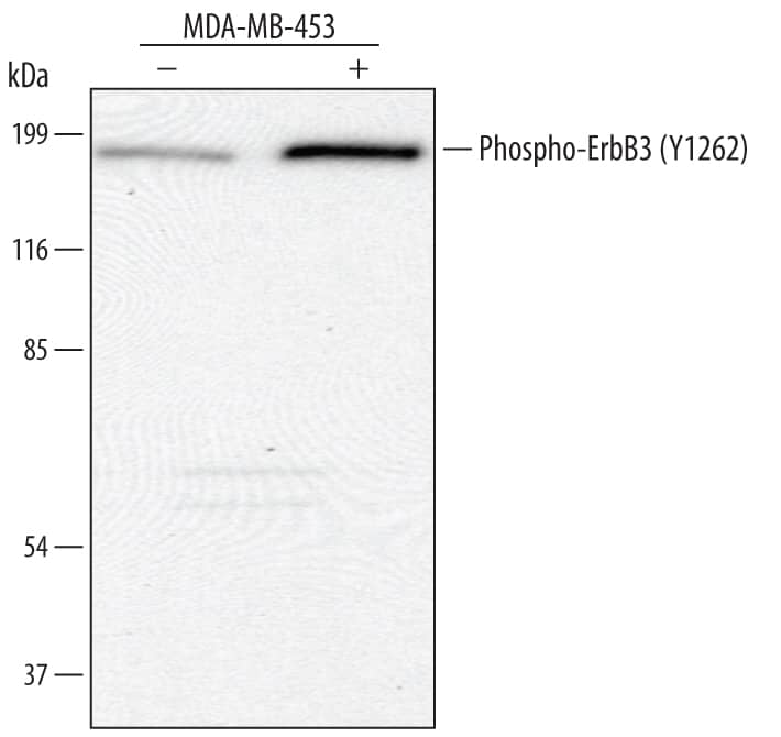
Detection of Human Phospho-ErbB3/Her3 (Y1262) by Western Blot.
Western blot shows lysates of MDA-MB-453 human breast cancer cell line untreated (-) or treated (+) with 300 ng/mL Recombinant Human NRG1-beta /HRG1-beta (Catalog # 396-HB) for 5 minutes. PVDF membrane was probed with 1 µg/mL of Human Phospho-ErbB3/Her3 (Y1262) Antigen Affinity-purified Polyclonal Antibody (Catalog # AF5817), followed by HRP-conjugated Anti-Rabbit IgG Secondary Antibody (Catalog # HAF008). A specific band was detected for Phospho-ErbB3/Her3 (Y1262) at approximately 185 kDa (as indicated). This experiment was conducted under reducing conditions and using Immunoblot Buffer Group 1.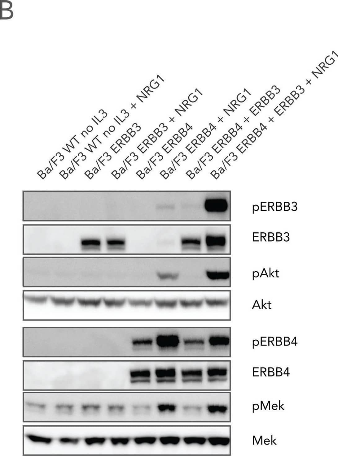
Detection of Phospho-ErbB3/Her3 (Y1262) by Western Blot
NRG1-induced proliferation and signaling of Ba/F3 cells expressing ERBB3 and/or ERBB4.(A) Cells expressing either ERBB4 (upper panel), ERBB4 and ERBB3 (middle panel) or ERBB3 receptors (lower panel) were cultured in medium without ligand, in the presence of 10 ng/ml IL3, or in the presence of 10 or 100 ng/ml NRG1. Cell numbers were counted at the indicated time points. (B) Parental Ba/F3 or Ba/F3 cells expressing ERBB3, ERBB4 or both receptors were incubated in the absence of IL3 and treated with 100 ng/mL NRG1 where indicated. Total cell lysate were then prepared and immunoblotted with phosphoepitope- and protein-specific antibodies for ERBB3, ERBB4, Akt and MEK. Image collected and cropped by CiteAb from the following open publication (https://pubmed.ncbi.nlm.nih.gov/26745281), licensed under a CC-BY license. Not internally tested by R&D Systems.Applications for Human Phospho-ErbB3/Her3 (Y1262) Antibody
Application
Recommended Usage
Western Blot
1 µg/mL
Sample: MDA-MB-453 human breast cancer cell line treated with Recombinant Human NRG1-beta /HRG1-beta (Catalog # 396-HB)
Sample: MDA-MB-453 human breast cancer cell line treated with Recombinant Human NRG1-beta /HRG1-beta (Catalog # 396-HB)
Formulation, Preparation, and Storage
Purification
Antigen Affinity-purified
Reconstitution
Reconstitute at 0.2 mg/mL in sterile PBS. For liquid material, refer to CoA for concentration.
Formulation
Lyophilized from a 0.2 μm filtered solution in PBS with Trehalose. *Small pack size (SP) is supplied either lyophilized or as a 0.2 µm filtered solution in PBS.
Shipping
Lyophilized product is shipped at ambient temperature. Liquid small pack size (-SP) is shipped with polar packs. Upon receipt, store immediately at the temperature recommended below.
Stability & Storage
Use a manual defrost freezer and avoid repeated freeze-thaw cycles.
- 12 months from date of receipt, -20 to -70 °C as supplied.
- 1 month, 2 to 8 °C under sterile conditions after reconstitution.
- 6 months, -20 to -70 °C under sterile conditions after reconstitution.
Background: ErbB3/Her3
ErbB3 (also known as HER3) is a type I transmembrane growth factor receptor and member of the human epidermal growth factor receptor (HER) family. ErbB3 is unique among HER family members in that it contains a defective kinase domain, and is therefore active only as a partner in a heterodimer complex with other ErbBs. Despite ErbB3's lack of intrinsic kinase activity, these heterodimers can be quite potent: the ErbB2/ErbB3 complex is considered the most active ErbB signaling dimer for the activation of downstream pathways. In addition, monomeric ErbB3 serves as a low affinity receptor for the heregulins (HRG), while ErbB2/ErbB3 heterodimers form a high affinity receptor complex. Phosphorylation of ErbB3 at Y1262 following ligand binding generates a binding site for the Grb2 adaptor protein.
Long Name
Receptor Tyrosine Protein Kinase ErbB3
Alternate Names
HER3
Gene Symbol
ERBB3
Additional ErbB3/Her3 Products
Product Documents for Human Phospho-ErbB3/Her3 (Y1262) Antibody
Product Specific Notices for Human Phospho-ErbB3/Her3 (Y1262) Antibody
For research use only
Loading...
Loading...
Loading...
Loading...
Loading...