
Human Phospho-Axl (Y779) Antibody
R&D Systems, part of Bio-Techne | Catalog # AF2228


Conjugate
Catalog #
Key Product Details
Species Reactivity
Validated:
Human
Cited:
Human, Mouse, Rat, Xenograft
Applications
Validated:
Immunocytochemistry
Cited:
Immunohistochemistry, Immunohistochemistry-Paraffin, Immunohistochemistry-Frozen, Western Blot, Flow Cytometry, Immunoprecipitation
Label
Unconjugated
Antibody Source
Polyclonal Rabbit IgG
Product Specifications
Immunogen
Phosphopeptide containing human Axl Y779 site
Specificity
Detects human and mouse Axl when phosphorylated at Y779.
Clonality
Polyclonal
Host
Rabbit
Isotype
IgG
Scientific Data Images for Human Phospho-Axl (Y779) Antibody
Phospho-Axl (Y779) in A172 Human Cell Line.
Axl phosphorylated at Y779 (panels B, D) and total Axl (panels A, C) were assessed in immersion fixed A172 human glioblastoma cells incubated with (panels C, D) or without (panels A, B) pervanadate. Phospho-Axl was detected using Rabbit Anti-Human Phospho-Axl (Y779) Antigen Affinity-purified Polyclonal Antibody (Catalog # AF2228) at 10 µg/mL for 3 hours at room temperature. Cells were stained using the NorthernLights™ 557-conjugated Anti-Rabbit IgG Secondary Antibody (red, panels B, D); NL004) and counterstained using DAPI (blue). Total Axl was detected using Goat Anti-Human Axl Antigen Affinity-purified Polyclonal Antibody (AF154). Cells were stained using the NorthernLights™ 493-conjugated Anti-Goat IgG Secondary Antibody (green, panels A, C); NL003). Specific staining was localized to cytoplasm. View our protocol for Fluorescent ICC Staining of Cells on Coverslips.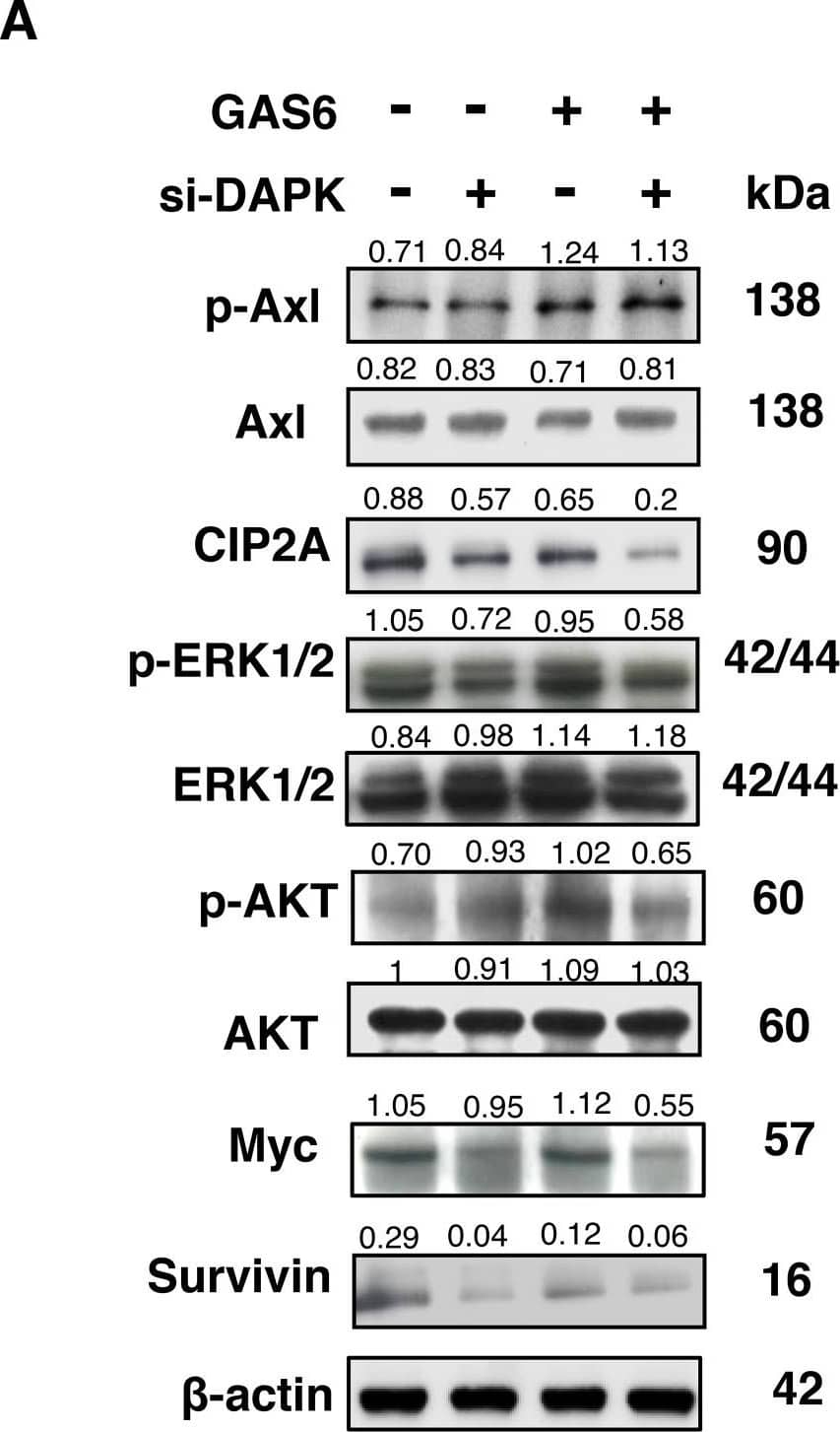
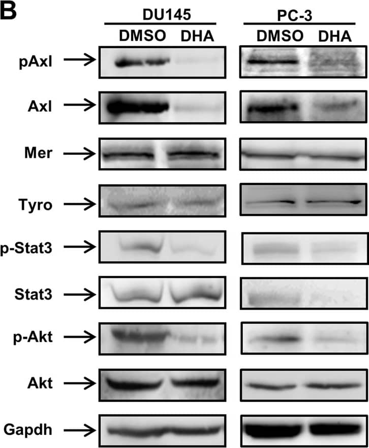
Detection of Rat Human Phospho-Axl (Y779) Antibody by Western Blot
GAS6 triggered Schwann cell proliferation primarily through CIP2A and DAPK(A) RSC96 cells were transfected with either control or DAPK siRNA for 48 h and then exposed to GAS6 (100 ng/ml) for 30 min. Immunoblotting evaluations of pAxl, Axl, CIP2A, DAPK, pERK1/2, ERK1/2, pAKT, AKT, Myc and Survivin. (B) RSC96 cells were transfected with either control or CIP2A siRNA for 48 h and then exposed to GAS6 (100 ng/ml) for 30 min. Immunoblotting evaluations of pAxl, Axl, DAPK, pDAPK, pERK1/2, ERK1/2, pAKT, AKT, Myc and Survivin. (C) DAPK or CIP2A was knocked down for 48 h then RSC96 cells were incubated with GAS6 (100 ng/ml) at the indicated hours. Cell viability was analysed via the WST-1 assay. Data are the mean ± SD, and n = 3 for each time point. * p < 0.05, ** p < 0.01 vs. scramble. Image collected and cropped by CiteAb from the following publication (https://pubmed.ncbi.nlm.nih.gov/29464081), licensed under a CC-BY license. Not internally tested by R&D Systems.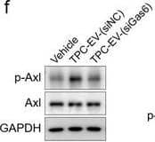
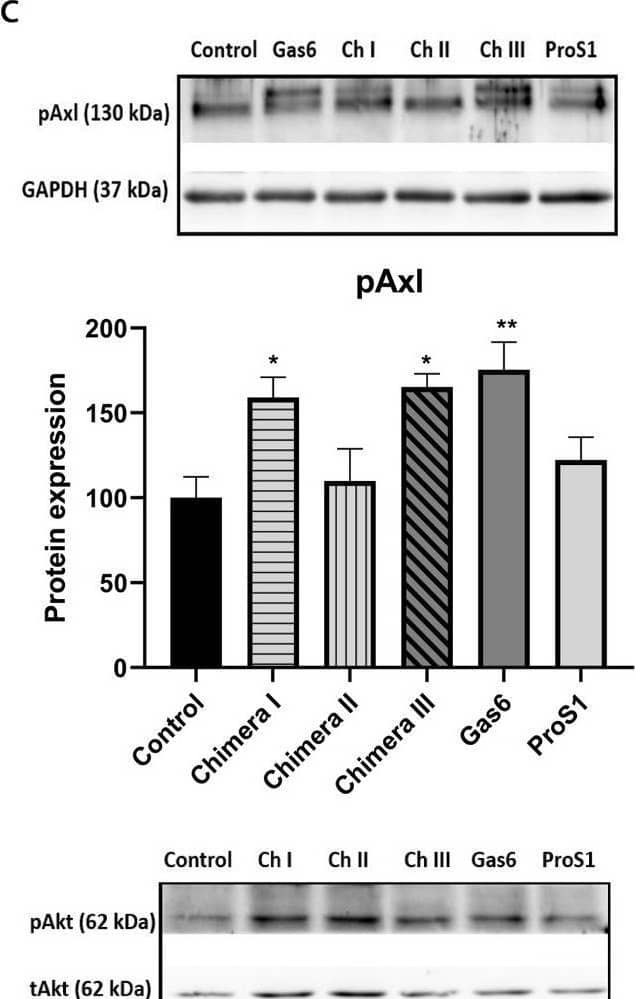
Detection of Human Axl by Western Blot
TPC‐EV‐induced EPC vasculogenic properties are regulated by Gas6 in vitro. (f) The phosphorylated&total forms of Axl, Akt&Erk1/2 in EPCs after the indicated treatments determined. Representative blots&quantification of the ratio of p‐Axl/Axl (n = 3). TPC‐EV‐(siNC), EVs derived from TPCs transfected with NC siRNA. TPC‐EV‐(siGas6), EVs derived from TPCs transfected with Gas6 siRNA. Image collected & cropped by CiteAb from the following open publication (https://pubmed.ncbi.nlm.nih.gov/34035882), licensed under a CC-BY license. Not internally tested by R&D Systems.
Applications for Human Phospho-Axl (Y779) Antibody
Application
Recommended Usage
Immunocytochemistry
5-15 µg/mL
Sample: Immersion fixed A172 human glioblastoma cell line with and without pervanadate co-staining with total Axl (Catalog # AF154)
Sample: Immersion fixed A172 human glioblastoma cell line with and without pervanadate co-staining with total Axl (Catalog # AF154)
Reviewed Applications
Read 6 reviews rated 4.2 using AF2228 in the following applications:
Formulation, Preparation, and Storage
Purification
Antigen and protein A Affinity-purified
Reconstitution
Reconstitute at 0.2 mg/mL in sterile PBS. For liquid material, refer to CoA for concentration.
Formulation
Lyophilized from a 0.2 μm filtered solution in PBS with Trehalose. See Certificate of Analysis for details.
*Small pack size (-SP) is supplied either lyophilized or as a 0.2 µm filtered solution in PBS.
*Small pack size (-SP) is supplied either lyophilized or as a 0.2 µm filtered solution in PBS.
Shipping
Lyophilized product is shipped at ambient temperature. Liquid small pack size (-SP) is shipped with polar packs. Upon receipt, store immediately at the temperature recommended below.
Stability & Storage
Use a manual defrost freezer and avoid repeated freeze-thaw cycles.
- 12 months from date of receipt, -20 to -70 °C as supplied.
- 1 month, 2 to 8 °C under sterile conditions after reconstitution.
- 6 months, -20 to -70 °C under sterile conditions after reconstitution.
Background: Axl
Axl (Ufo, Ark), Dtk (Sky, Tyro3, Rse, Brt), and Mer (human and mouse homologues of chicken c-Eyk) constitute a subfamily of the receptor tyrosine kinases (1, 2). The extracellular domains of these proteins contain two Ig-like motifs and two fibronectin type III motifs. This characteristic topology is also found in neural cell adhesion molecules and in receptor tyrosine phosphatases. The human Axl cDNA encodes an 887 amino acid (aa) precursor that includes an 18 aa signal sequence, a 426 aa extracellular domain, a 21 aa transmembrane segment, and a 422 aa cytoplasmic domain. The extracellular domains of human and mouse Axl share 81% aa sequence identity. A short alternately spliced form of human Axl is distinguished by a 9 aa deletion in the extracellular juxtamembrane region. These receptors bind the vitamin K‑dependent protein growth arrest specific gene 6 (Gas6) which is structurally related to the anticoagulation factor protein S. Binding of Gas6 induces receptor autophosphorylation and downstream signaling pathways that can lead to cell proliferation, migration, or the prevention of apoptosis (3). This family of tyrosine kinase receptors is involved in hematopoiesis, embryonic development, tumorigenesis, and regulation of testicular functions.
References
- Yanagita, M. (2004) Curr. Opin. Nephrol. Hypertens. 13:465.
- Nagata, K. et al. (1996) J. Biol. Chem. 22:30022.
- Holland, S. et al. (2005) Canc. Res. 65:9294.
Long Name
Axl Receptor Tyrosine Kinase
Alternate Names
AI323647, ARK, JTK11, Tyro7, UFO
Gene Symbol
AXL
Additional Axl Products
Product Documents for Human Phospho-Axl (Y779) Antibody
Product Specific Notices for Human Phospho-Axl (Y779) Antibody
For research use only
Loading...
Loading...
Loading...
Loading...
Loading...