Human Phospho-Axl (Y779) Antibody
R&D Systems, part of Bio-Techne | Catalog # MAB6965

Key Product Details
Validated by
Species Reactivity
Validated:
Cited:
Applications
Validated:
Cited:
Label
Antibody Source
Product Specifications
Immunogen
Specificity
Clonality
Host
Isotype
Scientific Data Images for Human Phospho-Axl (Y779) Antibody
Detection of Human Phospho-Axl (Y779) by Western Blot.
Western blot shows lysates of A172 human glioblastoma cell line untreated (-) or treated (+) with 1 mM Pervanadate (PV) for 30 minutes. PVDF membrane was probed with 0.2 µg/mL of Mouse Anti-Human Phospho-Axl (Y779) Monoclonal Antibody (Catalog # MAB6965) followed by HRP-conjugated Anti-Mouse IgG Secondary Antibody (Catalog # HAF007). A specific band was detected for Phospho-Axl (Y779) at approximately 140 kDa (as indicated). This experiment was conducted under reducing conditions and using Immunoblot Buffer Group 1.Phospho-Axl (Y779) in A172 Human Cell Line.
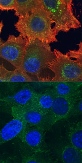
Axl phosphorylated at Y779 and total Axl were assessed in immersion fixed A172 human glioblastoma cells incubated with (upper panel) or without (lower panel) the phosphatase inhibitor pervanadate at 100 µM for 5 minutes. Phospho-Axl was detected using Mouse Anti-Human Phospho-Axl (Y779) Monoclonal Antibody (Catalog # MAB6965) at 10 µg/mL for 3 hours at room temperature. Cells were stained using the NorthernLights™ 557-conjugated Anti-Mouse IgG Secondary Antibody (red; Catalog # NL007) and counterstained using DAPI (blue). Total Axl was detected using Goat Anti-Human Axl Antigen Affinity-purified Polyclonal Antibody (Catalog # AF154). Cells were stained using the NorthernLights™ 493-conjugated Anti-Goat IgG Secondary Antibody (green; Catalog # NL003). Specific staining was localized to cytoplasm and cell surfaces. View our protocol for Fluorescent ICC Staining of Cells on Coverslips.Detection of Human Phospho-Axl (Y779) by Simple WesternTM.
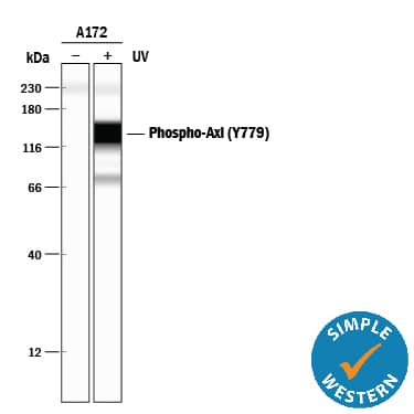
Simple Western lane view shows lysates of A172 human glioblastoma cell line untreated (-) or treated (+) with 1 mM Pervanadate (PV) for 30 minutes, loaded at 0.2 mg/mL. A specific band was detected for Phospho-Axl (Y779) at approximately 150 kDa (as indicated) using 4 µg/mL of Mouse Anti-Human Phospho-Axl (Y779) Monoclonal Antibody (Catalog # MAB6965). This experiment was conducted under reducing conditions and using the 12-230 kDa separation system.*Non-specific interaction with the 230 kDa Simple Western standard may be seen with this antibody.Applications for Human Phospho-Axl (Y779) Antibody
Immunocytochemistry
Sample: Immersion fixed A172 human glioblastoma cell line treated with or without pervanadate
Simple Western
Sample: A172 human glioblastoma cell line treated with Pervanadate (PV)
Western Blot
Sample: A172 human glioblastoma cell line treated with Pervanadate (PV)
Formulation, Preparation, and Storage
Purification
Reconstitution
Formulation
*Small pack size (-SP) is supplied either lyophilized or as a 0.2 µm filtered solution in PBS.
Shipping
Stability & Storage
- 12 months from date of receipt, -20 to -70 °C as supplied.
- 1 month, 2 to 8 °C under sterile conditions after reconstitution.
- 6 months, -20 to -70 °C under sterile conditions after reconstitution.
Background: Axl
Axl (Ufo, Ark), Dtk (Sky, Tyro3, Rse, Brt), and Mer (human and mouse homologues of chicken c-Eyk) constitute a subfamily of the receptor tyrosine kinases (1, 2). The extracellular domains of these proteins contain two Ig-like motifs and two fibronectin type III motifs. This characteristic topology is also found in neural cell adhesion molecules and in receptor tyrosine phosphatases. The human Axl cDNA encodes an 887 amino acid (aa) precursor that includes an 18 aa signal sequence, a 426 aa extracellular domain, a 21 aa transmembrane segment, and a 422 aa cytoplasmic domain. The extracellular domains of human and mouse Axl share 81% aa sequence identity. A short alternately spliced form of human Axl is distinguished by a 9 aa deletion in the extracellular juxtamembrane region. These receptors bind the vitamin K‑dependent protein growth arrest specific gene 6 (Gas6) which is structurally related to the anticoagulation factor protein S. Binding of Gas6 induces receptor autophosphorylation and downstream signaling pathways that can lead to cell proliferation, migration, or the prevention of apoptosis (3). This family of tyrosine kinase receptors is involved in hematopoiesis, embryonic development, tumorigenesis, and regulation of testicular functions. Phosphorylation of Tyrosine 779 provides a docking site for p85 subunits of PI 3-Kinase (4).
References
- Yanagita, M. (2004) Curr. Opin. Nephrol. Hypertens. 13:465.
- Nagata, K. et al. (1996) J. Biol. Chem. 22:30022.
- Holland, S. et al. (2005) Canc. Res. 65:9294.
- Weinger, J.G. et al. (2008) J. Neurochem. 106:134.
Long Name
Alternate Names
Gene Symbol
Additional Axl Products
Product Documents for Human Phospho-Axl (Y779) Antibody
Product Specific Notices for Human Phospho-Axl (Y779) Antibody
For research use only


