
Human Nbs1 Antibody
R&D Systems, part of Bio-Techne | Catalog # AF1573


Conjugate
Catalog #
Key Product Details
Validated by
Knockout/Knockdown
Species Reactivity
Human
Applications
Knockout Validated, Western Blot, Simple Western, Immunoprecipitation
Label
Unconjugated
Antibody Source
Polyclonal Goat IgG
Product Specifications
Immunogen
E. coli-derived recombinant human Nbs1
Pro498-Arg754
Accession # O60934
Pro498-Arg754
Accession # O60934
Specificity
Detects human Nbs1 in Western blots.
Clonality
Polyclonal
Host
Goat
Isotype
IgG
Scientific Data Images for Human Nbs1 Antibody
Detection of Human Nbs1 by Western Blot.
Western blot shows lysates of HeLa human cervical epithelial carcinoma cell line and K562 human chronic myelogenous leukemia cell line. PVDF membrane was probed with 0.5 µg/mL of Goat Anti-Human Nbs1 Antigen Affinity-purified Polyclonal Antibody (Catalog # AF1573) followed by HRP-conjugated Anti-Goat IgG Secondary Antibody (Catalog # HAF017). A specific band was detected for Nbs1 at approximately 100 kDa (as indicated). This experiment was conducted under reducing conditions and using Immunoblot Buffer Group 1.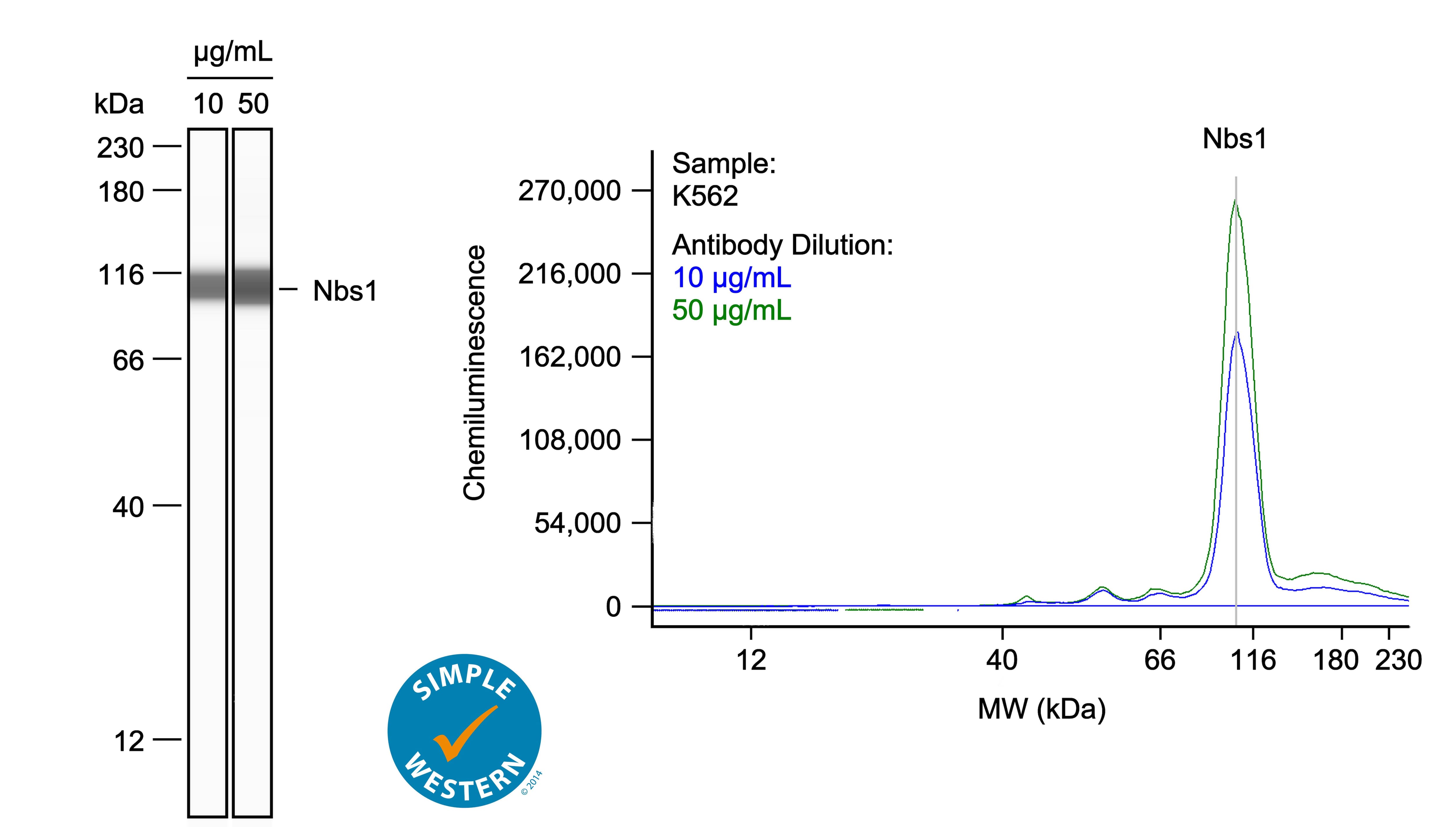
Detection of Human Nbs1 by Simple WesternTM.
Left: Simple Western lane view shows lysates of K562 human chronic myelogenous leukemia cell line, loaded at 0.1 mg/ml. A specific band was detected for Nbs1 at approximately 109 kDa (as indicated) using both 10 µg/ml and 50 µg/ml of Goat Anti-Human Nbs1 Antigen Affinity-purified Polyclonal Antibody (Catalog # AF1573) followed by HRP-conjugated Donkey Anti-Goat Secondary Antibody (Catalog # 043-522-2). This experiment was conducted under reducing conditions and using the 12-230kDa separation system. Right: Simple Western electropherogram showing the same Goat Anti-Human Nbs1 Antigen Affinity-purified Polyclonal Antibody (Catalog # AF1573) tested at 10 µg/ml (blue line) and 50 µg/ml (green line) in the K562 human chronic myelogenous leukemia cell line.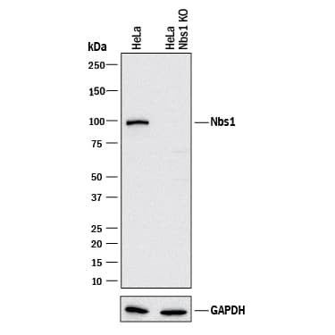
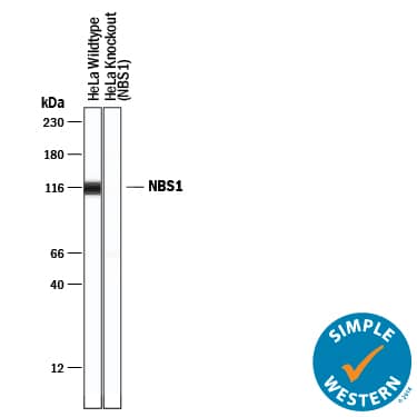
Western Blot Shows Human Nbs1 Specificity by Using Knockout Cell Line.
Western blot shows lysates of HeLa human cervical epithelial carcinoma parental cell line and Nbs1 knockout HeLa cell line (KO). PVDF membrane was probed with 0.25 µg/mL of Goat Anti-Human Nbs1 Antigen Affinity-purified Polyclonal Antibody (Catalog # AF1573) followed by HRP-conjugated Anti-Goat IgG Secondary Antibody (Catalog # HAF017). A specific band was detected for Nbs1 at approximately 95 kDa (as indicated) in the parental HeLa cell line, but is not detectable in knockout HeLa cell line. GAPDH (Catalog # AF5718) is shown as a loading control. This experiment was conducted under reducing conditions and using Immunoblot Buffer Group 1.Applications for Human Nbs1 Antibody
Application
Recommended Usage
Immunoprecipitation
2 µg/500 µg cell lysate
Sample: HeLa human cervical epithelial carcinoma cell line and K562 human chronic myelogenous leukemia cell line, see our available Western blot detection antibodies
Sample: HeLa human cervical epithelial carcinoma cell line and K562 human chronic myelogenous leukemia cell line, see our available Western blot detection antibodies
Knockout Validated
Nbs1
is specifically detected in HeLa human cervical epithelial carcinoma parental cell line but is not detectable in
Nbs1 knockout HeLa cell line.
Simple Western
10-50 µg/mL
Sample: K562 human chronic myelogenous leukemia cell line, HeLa human cervical epithelial carcinoma cell line
Sample: K562 human chronic myelogenous leukemia cell line, HeLa human cervical epithelial carcinoma cell line
Western Blot
0.5 µg/mL
Sample: HeLa human cervical epithelial carcinoma cell line and K562 human chronic myelogenous leukemia cell line
Sample: HeLa human cervical epithelial carcinoma cell line and K562 human chronic myelogenous leukemia cell line
Formulation, Preparation, and Storage
Purification
Antigen Affinity-purified
Reconstitution
Reconstitute at 0.2 mg/mL in sterile PBS. For liquid material, refer to CoA for concentration.
Formulation
Lyophilized from a 0.2 μm filtered solution in PBS with Trehalose. See Certificate of Analysis for details.
*Small pack size (-SP) is supplied either lyophilized or as a 0.2 µm filtered solution in PBS.
*Small pack size (-SP) is supplied either lyophilized or as a 0.2 µm filtered solution in PBS.
Shipping
Lyophilized product is shipped at ambient temperature. Liquid small pack size (-SP) is shipped with polar packs. Upon receipt, store immediately at the temperature recommended below.
Stability & Storage
Use a manual defrost freezer and avoid repeated freeze-thaw cycles.
- 12 months from date of receipt, -20 to -70 °C as supplied.
- 1 month, 2 to 8 °C under sterile conditions after reconstitution.
- 6 months, -20 to -70 °C under sterile conditions after reconstitution.
Background: Nbs1
The Nijmegen Breakage Syndrome 1 (Nbs1) protein is a member of the Mre11/Rad50/Nbs1 (MRN) protein complex that binds to DNA double-strand breaks in cells exposed to DNA damaging agents. In addition, the MRN complex colocalizes with replication forks during DNA replication. The MRN complex plays an important role in routine cell cycle progression and genotoxic stress responses by facilitating DNA repair. In fact, mutation of the nbs1 gene and resultant loss of Nbs1 protein expression in humans results in the chromosomal instability disease, Nijmegen Breakage Syndrome.
Long Name
Nijmegen Breakage Syndrome 1
Alternate Names
NBN, Nibrin, p95
Gene Symbol
NBN
UniProt
Additional Nbs1 Products
Product Documents for Human Nbs1 Antibody
Product Specific Notices for Human Nbs1 Antibody
For research use only
Loading...
Loading...
Loading...
Loading...
Loading...