
Human/Mouse NeuroD1 Antibody
R&D Systems, part of Bio-Techne | Catalog # AF2746

Scientific images may be resized, cropped, or adjusted for brightness or contrast for presentation purposes. These updates are limited to presentation and do not affect the underlying scientific interpretation of the data. Where available, additional source data may be provided upon request.
Conjugate
Catalog #
Key Product Details
Species Reactivity
Validated:
Human, Mouse
Cited:
Human, Mouse, Rat, Transgenic Mouse, Xenograft
Applications
Validated:
Western Blot, Immunocytochemistry
Cited:
Immunohistochemistry, Immunohistochemistry-Paraffin, Flow Cytometry, Immunocytochemistry
Label
Unconjugated
Antibody Source
Polyclonal Goat IgG
Product Specifications
Immunogen
E. coli-derived recombinant human NeuroD1
Ser154-Asp356
Accession # Q13562
Ser154-Asp356
Accession # Q13562
Specificity
Detects NeuroD1 in direct ELISAs and Western blots. In these formats, approximately 5% cross-reactivity with recombinant human NeuroD2 is observed.
Clonality
Polyclonal
Host
Goat
Isotype
IgG
Scientific Data Images for Human/Mouse NeuroD1 Antibody
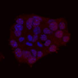
NeuroD1 in betaTC-6 Mouse Cell Line.
Neurogenic Differentiation factor 1 (NeuroD1) was detected in immersion fixed betaTC-6 mouse beta cell insulinoma cell line using Human/Mouse NeuroD1 Antigen Affinity-purified Polyclonal Antibody (Catalog # AF2746) at 10 µg/mL for 3 hours at room temperature. Cells were stained using the NorthernLights™ 557-conjugated Anti-Goat IgG Secondary Antibody (red; Catalog # NL001) and counterstained with DAPI (blue). View our protocol for Fluorescent ICC Staining of Cells on Coverslips.
Detection of NeuroD1 by Immunohistochemistry
Organoid characterization(A) Representative bright-field image of an organoid on the seventh day of culture.(B and C) Immunostaining of a representative organoid for iOSNs (Gap43), HBCs (KRT5), and GBCs (SOX2+, KRT5-). (C) shows high-magnification images of (B).(D–F) Representative immunostaining of organoids for other neurogenic markers.(G) UMAP representation of single-cell RNA-seq of the organoid culture.(H) Stacked violin plots of key marker genes allowing cluster annotation in log scale.(I) Focused UMAP expression plots in the dotted area shown in (G), plotted in log-scale, representing the neurogenic cascade found in the organoids.(J) Histogram plot of the number of distinct ORs found in single cells identified as OSNs based on UCHL1, GAP43, and TUBB3. Image collected and cropped by CiteAb from the following open publication (https://linkinghub.elsevier.com/retrieve/pii/S2667237525000918), licensed under a CC-BY license. Not internally tested by R&D Systems.
Detection of NeuroD1 by Immunohistochemistry
MCT8-deficient COs present an altered transcriptome.(A) Schematic of the protocol for COs’ maturation using platelet-derived growth factor AA and insulin-like growth factor 1. (B–E) Whole-mount immunolabeling of D65 WT COs containing differentiated NEUROD1+ neurons (B), GFAP+ neural precursor cells and GFAP-AQP4+ astrocytes (C and D), and OLIG2+ oligodendrocyte precursor cells and O4+ premyelinating oligodendrocytes (E); in blue is the nuclear staining with DAPI. B: scale bar 75 μm; C: scale bar 75 μm; inset in C: scale bar 25 μm; D: scale bar 40 μm; E: scale bar 75 μm; inset in E: scale bar 40 μm. Arrows in D indicate AQP4 staining in the membrane of GFAP+ cells. The arrow in the inset in E points to the cellular body of a representative oligodendrocyte; arrowheads point to its cellular processes. (F and G) Volcano plots showing the distribution of DEGs in WT vs. Mut1 (F) and in WT vs. Mut2 (G); each point represents the average of 3 (WT) or 2 (MCT8-deficient) samples consisting of 4 pooled COs for each transcript. (H) Venn comparison of WT vs. Mut1, WT vs. Mut2, and Mut1 vs. Mut2 DEGs. A common cluster (cluster E) between WT vs. Mut1 and WT vs. Mut2 containing 949 DEGs was identified. (I) Venn comparison of the 949 DEGs in cluster E shows similar transcriptomic changes in both MCT8-deficient COs. (J) Heatmap depicting the top 63 DEGs from cluster E. (K) KEGG pathway analysis of the top 63 DEGs in J. (L–O) Heatmaps of T3-regulated genes involved in cerebral cortex development (L), neural cell migration (M), astrocytes and myelination (N), and neurotransmitter receptors, transcription factors, potassium channels, and extracellular matrix protein (O). DEG thresholds (FDR ≤ 0.25 and fold-change ± 4) were identified by gene set–specific analysis in the Partek Flow platform. Image collected and cropped by CiteAb from the following open publication (https://pubmed.ncbi.nlm.nih.gov/38376950), licensed under a CC-BY license. Not internally tested by R&D Systems.Applications for Human/Mouse NeuroD1 Antibody
Application
Recommended Usage
Immunocytochemistry
5-15 µg/mL
Sample: Immersion fixed beta TC-6 mouse beta cell insulinoma cell line
Sample: Immersion fixed beta TC-6 mouse beta cell insulinoma cell line
Western Blot
0.1 µg/mL
Sample: Recombinant Human NeuroD1
Sample: Recombinant Human NeuroD1
Reviewed Applications
Read 1 review rated 5 using AF2746 in the following applications:
Formulation, Preparation, and Storage
Purification
Antigen Affinity-purified
Reconstitution
Reconstitute at 0.2 mg/mL in sterile PBS. For liquid material, refer to CoA for concentration.
Formulation
Lyophilized from a 0.2 μm filtered solution in PBS with Trehalose. *Small pack size (SP) is supplied either lyophilized or as a 0.2 µm filtered solution in PBS.
Shipping
Lyophilized product is shipped at ambient temperature. Liquid small pack size (-SP) is shipped with polar packs. Upon receipt, store immediately at the temperature recommended below.
Stability & Storage
Use a manual defrost freezer and avoid repeated freeze-thaw cycles.
- 12 months from date of receipt, -20 to -70 °C as supplied.
- 1 month, 2 to 8 °C under sterile conditions after reconstitution.
- 6 months, -20 to -70 °C under sterile conditions after reconstitution.
Background: NeuroD1
Long Name
Neurogenic Differentiation Factor 1
Alternate Names
BETA2, BHF-1, NIDDM
Entrez Gene IDs
4760 (Human)
Gene Symbol
NEUROD1
UniProt
Additional NeuroD1 Products
Product Documents for Human/Mouse NeuroD1 Antibody
Product Specific Notices for Human/Mouse NeuroD1 Antibody
For research use only
Loading...
Loading...
Loading...
Loading...
Loading...