
Human/Mouse Histone Deacetylase 8/HDAC8 Antibody
R&D Systems, part of Bio-Techne | Catalog # AF4359


Scientific images may be resized, cropped, or adjusted for brightness or contrast for presentation purposes. These updates are limited to presentation and do not affect the underlying scientific interpretation of the data. Where available, additional source data may be provided upon request.
Conjugate
Catalog #
Key Product Details
Validated by
Biological Validation
Species Reactivity
Human, Mouse
Applications
Western Blot, Chromatin Immunoprecipitation (ChIP)
Label
Unconjugated
Antibody Source
Polyclonal Sheep IgG
Product Specifications
Immunogen
S. frugiperda insect ovarian cell line Sf 21-derived recombinant human HDAC8 isoform 1
Met1-Val377 (Leu31Pro)
Accession # Q9BY41
Met1-Val377 (Leu31Pro)
Accession # Q9BY41
Specificity
Detects human HDAC8 in direct ELISAs and Western blots.
Clonality
Polyclonal
Host
Sheep
Isotype
IgG
Scientific Data Images for Human/Mouse Histone Deacetylase 8/HDAC8 Antibody
Detection of Human Histone Deacetylase 8/HDAC8 by Western Blot.
Western blot shows lysates of HEK293 human embryonic kidney cell line. PVDF membrane was probed with 1 µg/mL of Sheep Anti-Human/Mouse Histone Deacetylase 8/HDAC8 Antigen Affinity-purified Polyclonal Antibody (Catalog # AF4359) followed by HRP-conjugated Anti-Sheep IgG Secondary Antibody (Catalog # HAF016). A specific band was detected for Histone Deacetylase 8/HDAC8 at approximately 50 kDa (as indicated). This experiment was conducted under reducing conditions and using Immunoblot Buffer Group 8.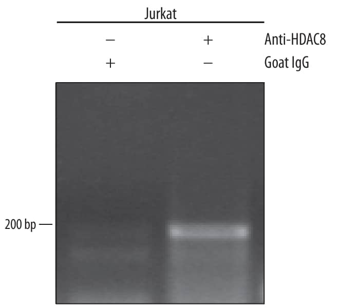
Detection of Histone Deacetylase 8/ HDAC8-regulated Genes by Chromatin Immunoprecipitation.
Jurkat human acute T cell leukemia cell line treated with 50 ng/mL PMA and 200 ng/mL calcium ionomycin for 30 minutes was fixed using formaldehyde, resuspended in lysis buffer, and sonicated to shear chromatin. Histone Deacetylase 8/HDAC8/DNA complexes were immunoprecipitated using 5 µg Sheep Anti-Human/Mouse Histone Deacetylase 8/HDAC8 Antigen Affinity-purified Polyclonal Antibody (Catalog # AF4359) or control antibody (Catalog # 5-001-A) for 15 minutes in an ultrasonic bath, followed by Biotinylated Anti-Sheep IgG Secondary Antibody (Catalog # BAF016).Immunocomplexes were captured using 50 µL of MagCellect Streptavidin Ferrofluid (Catalog # MAG999) and DNA was purified using chelating resin solution. TheIFN-gammapromoter was detected by standard PCR.Applications for Human/Mouse Histone Deacetylase 8/HDAC8 Antibody
Application
Recommended Usage
Chromatin Immunoprecipitation (ChIP)
5 µg/5 x 106 cells
Sample: PMA and calcium ionomycin treated Jurkat human acute T cell leukemia cell line chromatin, IFN-gamma promoter detected by standard PCR
Sample: PMA and calcium ionomycin treated Jurkat human acute T cell leukemia cell line chromatin, IFN-gamma promoter detected by standard PCR
Western Blot
1 µg/mL
Sample: HEK293 human embryonic kidney cell line
Sample: HEK293 human embryonic kidney cell line
Formulation, Preparation, and Storage
Purification
Antigen Affinity-purified
Reconstitution
Reconstitute at 0.2 mg/mL in sterile PBS. For liquid material, refer to CoA for concentration.
Formulation
Lyophilized from a 0.2 μm filtered solution in PBS with Trehalose. *Small pack size (SP) is supplied either lyophilized or as a 0.2 µm filtered solution in PBS.
Shipping
Lyophilized product is shipped at ambient temperature. Liquid small pack size (-SP) is shipped with polar packs. Upon receipt, store immediately at the temperature recommended below.
Stability & Storage
Use a manual defrost freezer and avoid repeated freeze-thaw cycles.
- 12 months from date of receipt, -20 to -70 °C as supplied.
- 1 month, 2 to 8 °C under sterile conditions after reconstitution.
- 6 months, -20 to -70 °C under sterile conditions after reconstitution.
Background: Histone Deacetylase 8/HDAC8
HDAC8 (Histone deacetylase 8) is a 50 kDa member of the class I subfamily, histone deacetylase family of transcriptional regulators. It is a smooth muscle enzyme that deacetylates histone lysines, blocking gene transcription. HDAC8 appears to function as a monomer, and phosphorylation at Ser39 inhibits activity. Human HDAC8 is 377 amino acids (aa) in length. It contains a series of alpha helices and strands, plus a catalytic ferrous ion binding site (Asp178-His180-Asp267). There are four potential splice variants, all of which possess an alternate start site 38 aa upstream of the standard start site. Three have additional changes; two show a one and three aa substitution respectively for aa 211-377, while a third has a 15 aa substitution for aa 148-377. Human HDAC8 shares more than 96% aa identity with both mouse and canine HDAC8.
Alternate Names
HDAC8, HDACL1, RPD3
Gene Symbol
HDAC8
UniProt
Additional Histone Deacetylase 8/HDAC8 Products
Product Documents for Human/Mouse Histone Deacetylase 8/HDAC8 Antibody
Product Specific Notices for Human/Mouse Histone Deacetylase 8/HDAC8 Antibody
For research use only
Loading...
Loading...
Loading...
Loading...
Loading...