
Human Mitofusin 1 Antibody
R&D Systems, part of Bio-Techne | Catalog # AF7880

Scientific images may be resized, cropped, or adjusted for brightness or contrast for presentation purposes. These updates are limited to presentation and do not affect the underlying scientific interpretation of the data. Where available, additional source data may be provided upon request.
Conjugate
Catalog #
Key Product Details
Species Reactivity
Validated:
Human
Cited:
Human, Rat
Applications
Validated:
Western Blot
Cited:
Western Blot
Label
Unconjugated
Antibody Source
Polyclonal Sheep IgG
Product Specifications
Immunogen
E. coli-derived recombinant human Mitofusin 1
Ala2-Lys77
Accession # Q8IWA4
Ala2-Lys77
Accession # Q8IWA4
Specificity
Detects human Mitofusin 1 in direct ELISAs and Western blots. In direct ELISAs, less than 3% cross-reactivity with recombinant human Mitofusin 2 is observed.
Clonality
Polyclonal
Host
Sheep
Isotype
IgG
Scientific Data Images for Human Mitofusin 1 Antibody
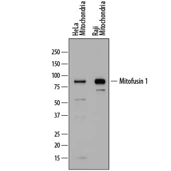
Detection of Human Mitofusin 1 by Western Blot.
Western blot shows mitochondrial extracts of HeLa human cervical epithelial carcinoma cell line (20 µg/lane) and Raji human Burkitt's lymphoma cell line (2.5 µg/lane). PVDF membrane was probed with 1 µg/mL of Sheep Anti-Human Mitofusin 1 Antigen Affinity-purified Polyclonal Antibody (Catalog # AF7880) followed by HRP-conjugated Anti-Sheep IgG Secondary Antibody (Catalog # HAF016). A specific band was detected for Mitofusin 1 at approximately 85 kDa (as indicated). This experiment was conducted under reducing conditions and using Immunoblot Buffer Group 1.Applications for Human Mitofusin 1 Antibody
Application
Recommended Usage
Western Blot
1 µg/mL
Sample: Mitochondrial extracts of HeLa human cervical epithelial carcinoma cell line and Raji human Burkitt's lymphoma cell line
Sample: Mitochondrial extracts of HeLa human cervical epithelial carcinoma cell line and Raji human Burkitt's lymphoma cell line
Formulation, Preparation, and Storage
Purification
Antigen Affinity-purified
Reconstitution
Sterile PBS to a final concentration of 0.2 mg/mL. For liquid material, refer to CoA for concentration.
Formulation
Lyophilized from a 0.2 μm filtered solution in PBS with Trehalose. *Small pack size (SP) is supplied either lyophilized or as a 0.2 µm filtered solution in PBS.
Shipping
Lyophilized product is shipped at ambient temperature. Liquid small pack size (-SP) is shipped with polar packs. Upon receipt, store immediately at the temperature recommended below.
Stability & Storage
Use a manual defrost freezer and avoid repeated freeze-thaw cycles.
- 12 months from date of receipt, -20 to -70 °C as supplied.
- 1 month, 2 to 8 °C under sterile conditions after reconstitution.
- 6 months, -20 to -70 °C under sterile conditions after reconstitution.
Background: Mitofusin 1
MFN-1 (Mitofusin 1; also Fzo/"fuzzy onions" homolog and Transmembrane GTPase MFN-1) is an 80-90 kDa mitochondrial member of the dynamin family of molecules. It is ubiquitously expressed, and found in the outer mitochondrial membrane. MFN-1 apparently mediates the fusion of individual mitochondria with each other through trans-interactions with adjacent mitochondrial MFN-1 and/or MFN-2. Pertubations with this process result in both a general overall fragmentation of mitochondria, and in cell-specific effects such as a reduction in oxidative phosphorylation or an imbalance in the generation of ROS. MFN-1 has two key domains. One is a coiled-coil region that mediates MFN-1:MFN-1/2 binding, and a second is a GTPase domain whose cleavage of GTP is necessary for membrane fusion. Human MFN-1 is a 741 amino acid (aa) two transmembrane protein that contains a cytoplasmic N- and C-terminus. The cytoplasmic N-terminus (aa 1-584) contains the GTPase domain (aa 82-286) while the cytoplasmic C-terminus possesses a critical coiled-coil motif (aa 679-734). MFN-1 is known to form oligomers, either with itself or MFN-2, and to undergo ubiquitination by MARCH5. Multiple splice variants are reported to occur. One is cytosolic and contains a four aa substitution for aa 367-741. Three others show a deletion of aa 444-554, aa 326-408 and an alternative start site at Met387, respectively. Over aa 1-77, human and mouse MFN-1 share 88% aa sequence identity. Full-length human MFN-1 and MFN-2 share 61% aa sequence identity.
Alternate Names
hfzo1, hfzo2, MFN1
Gene Symbol
MFN1
UniProt
Additional Mitofusin 1 Products
Product Documents for Human Mitofusin 1 Antibody
Product Specific Notices for Human Mitofusin 1 Antibody
For research use only
Loading...
Loading...
Loading...
Loading...
Loading...