Human IL-16 C-terminal Peptide Antibody
R&D Systems, part of Bio-Techne | Catalog # AF-316-PB


Key Product Details
Species Reactivity
Validated:
Cited:
Applications
Validated:
Cited:
Label
Antibody Source
Product Specifications
Immunogen
Met1203-Ser1332
Accession # Q14005
Specificity
Clonality
Host
Isotype
Scientific Data Images for Human IL-16 C-terminal Peptide Antibody
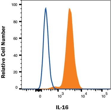
Detection of IL-16 in Raji cells by Flow Cytometry
Raji cells were stained with Goat Anti-Human IL-16 C-terminal Peptide Antigen Affinity-purified Polyclonal Antibody (Catalog # AF-316-PB, filled histogram) or isotype control antibody (Catalog # AB-108-C, open histogram) followed by Allophycocyanin-conjugated Anti-Goat IgG Secondary Antibody (Catalog # F0108). To facilitate intracellular staining, cells were fixed with Flow Cytometry Fixation Buffer (Catalog # FC004) and permeabilized with Flow Cytometry Permeabilization/Wash Buffer I (Catalog # FC005). View our protocol for Staining Intracellular Molecules.Detection of Human IL‑16 by Western Blot.
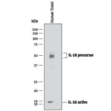
Western blot shows lysate of human tonsil tissue. PVDF membrane was probed with 1 µg/mL of Goat Anti-Human IL-16 C-terminal Peptide Antigen Affinity-purified Polyclonal Antibody (Catalog # AF-316-PB) followed by HRP-conjugated Anti-Goat IgG Secondary Antibody (Catalog # HAF017). Specific bands were detected for IL-16 at approximately 14 kDa (active) and 45-55 kDa (precursor), as indicated. This experiment was conducted under reducing conditions and using Immunoblot Buffer Group 1.IL-16 in Human PBMCs.
IL-16 was detected in immersion fixed LPS-stimulated human peripheral blood mononuclear cells (PBMCs) using 10 µg/mL Human IL-16 C-terminal Peptide Antigen Affinity-purified Polyclonal Antibody (Catalog # AF-316-PB) for 3 hours at room temperature. Cells were stained with the NorthernLights™ 557-conjugated Anti-Goat IgG Secondary Antibody (red; Catalog # NL001) and counterstained with DAPI (blue). View our protocol for Fluorescent ICC Staining of Non-adherent Cells.Applications for Human IL-16 C-terminal Peptide Antibody
CyTOF-ready
Immunocytochemistry
Sample: Immersion fixed human peripheral blood mononuclear cells (PBMCs) treated with PHA and immersion fixed human PBMCs treated LPS
Immunohistochemistry
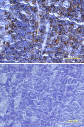
Sample: Immersion fixed paraffin-embedded sections of human tonsil
Intracellular Staining by Flow Cytometry
Sample: Raji human Burkitt's lymphoma cell line fixed with paraformaldehyde and permeabilized with saponin
Western Blot
Sample: Human tonsil tissue
Reviewed Applications
Read 2 reviews rated 5 using AF-316-PB in the following applications:
Formulation, Preparation, and Storage
Purification
Reconstitution
Formulation
*Small pack size (-SP) is supplied either lyophilized or as a 0.2 µm filtered solution in PBS.
Shipping
Stability & Storage
- 12 months from date of receipt, -20 to -70 °C as supplied.
- 1 month, 2 to 8 °C under sterile conditions after reconstitution.
- 6 months, -20 to -70 °C under sterile conditions after reconstitution.
Background: IL-16
Interleukin 16, also named lymphocyte chemoattractant factor (LCF), was originally identified as a CD8+ T-cell-derived chemoattractant for CD4+ cells. The biologically active form of IL-16 was originally proposed to be a homotetramer of 14 kDa chains containing 130 amino acid residue subunits. The complete pro-IL-16 cDNA was subsequently cloned and shown to encode a 631 amino acid residue hydrophilic protein that lacked a signal peptide. The original 130 amino acid residue polypeptide is now believed to have been derived from the C terminus of the precursor. IL-16 precursor protein has been detected in the lysates of various cells including mitogen stimulated PBMCs. The biologically active and secreted natural IL-16 is assumed to be a proteolytic cleavage product of pro-IL-16 generated by proteases present in or on activated CD8+ cells. A likely cleavage site was proposed to be at aspartate residue 510. This would yield a 121 amino acid residue protein, smaller than the 130 aa residue protein first described. The expression of IL-16 precursor mRNA has been detected in various tissues including spleen, thymus, lymph nodes, peripheral leukocytes, bone marrow and cerebellum. The gene for IL-16 precursor has been localized to chromosome 15. The biological activities ascribed to IL-16 are reported to be dependent on the cell surface expression of CD4, suggesting that IL-16 is a CD4 ligand. Besides its chemotactic properties, IL-16 has also been shown to suppress HIV-1 replication in vitro. Recombinant E. coli-derived IL-16 produced at R&D Systems is present mostly as a monomer, exhibits chemotactic activity for lymphocytes at high concentrations, lacks chemotactic activites for monocytes, and binds the extracellular domain of CD4 with low affinity.
References
- Cruikshank, W.W. et al. (1994) Proc. Natl. Acad. Sci. USA 91:5109.
- Baier, M. et al. (1997) Proc. Natl. Acad. Sci. USA 94:5273.
- Zhou, A. et al. (1997) Nature Medicine 3:659.
- Bazan, J.F. and T.J. Schall (1996) Nature 381:29.
Long Name
Alternate Names
Gene Symbol
UniProt
Additional IL-16 Products
Product Documents for Human IL-16 C-terminal Peptide Antibody
Product Specific Notices for Human IL-16 C-terminal Peptide Antibody
For research use only


