
Human ICAM-4 Antibody
R&D Systems, part of Bio-Techne | Catalog # AF7179


Scientific images may be resized, cropped, or adjusted for brightness or contrast for presentation purposes. These updates are limited to presentation and do not affect the underlying scientific interpretation of the data. Where available, additional source data may be provided upon request.
Conjugate
Catalog #
Key Product Details
Species Reactivity
Human
Applications
Western Blot, Flow Cytometry, Simple Western, CyTOF-ready
Label
Unconjugated
Antibody Source
Polyclonal Sheep IgG
Product Specifications
Immunogen
Chinese hamster ovary cell line CHO-derived recombinant human ICAM-4
Ala31-Ala240
Accession # Q14773
Ala31-Ala240
Accession # Q14773
Specificity
Detects human ICAM-4 in direct ELISAs and Western blots.
Clonality
Polyclonal
Host
Sheep
Isotype
IgG
Scientific Data Images for Human ICAM-4 Antibody
Detection of Human ICAM‑4 by Western Blot.
Western blot shows lysates of human erythrocytes and human heart tissue. PVDF membrane was probed with 0.2 µg/mL of Sheep Anti-Human ICAM-4 Antigen Affinity-purified Polyclonal Antibody (Catalog # AF7179) followed by HRP-conjugated Anti-Sheep IgG Secondary Antibody (Catalog # HAF016). A specific band was detected for ICAM-4 at approximately 42 kDa (as indicated). This experiment was conducted under reducing conditions and using Immunoblot Buffer Group 1.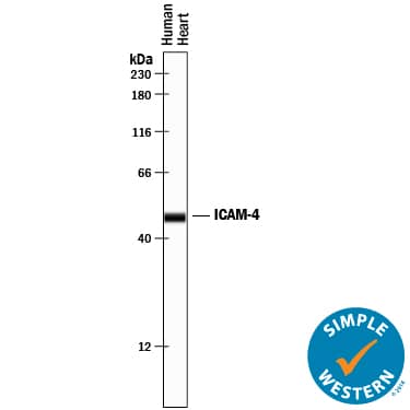
Detection of Human ICAM-4 by Simple WesternTM.
Simple Western lane view shows lysates of human heart tissue, loaded at 0.2 mg/mL. A specific band was detected for ICAM-4 at approximately 48 kDa (as indicated) using 10 µg/mL of Sheep Anti-Human ICAM-4 Antigen Affinity-purified Polyclonal Antibody (Catalog # AF7179) followed by 1:50 dilution of HRP-conjugated Anti-Sheep IgG Secondary Antibody (Catalog # HAF016). This experiment was conducted under reducing conditions and using the 12-230 kDa separation system.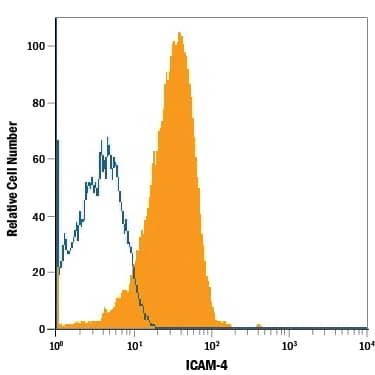
Detection of ICAM-4 in Human Red Blood Cells by Flow Cytometry.
Human red blood cells (erythrocytes) were stained with Sheep Anti-Human ICAM-4 Antigen Affinity-purified Polyclonal Antibody (Catalog # AF7179, filled histogram) or isotype control antibody (Catalog # 5-001-A, open histogram), followed by Allophycocyanin-conjugated Anti-Sheep IgG Secondary Antibody (Catalog # F0127).Applications for Human ICAM-4 Antibody
Application
Recommended Usage
CyTOF-ready
Ready to be labeled using established conjugation methods. No BSA or other carrier proteins that could interfere with conjugation.
Flow Cytometry
2.5 µg/106 cells
Sample: Human red blood cells (erythrocytes)
Sample: Human red blood cells (erythrocytes)
Simple Western
10 µg/mL
Sample: Human heart tissue
Sample: Human heart tissue
Western Blot
0.2 µg/mL
Sample: Human erythrocytes and human heart tissue
Sample: Human erythrocytes and human heart tissue
Formulation, Preparation, and Storage
Purification
Antigen Affinity-purified
Reconstitution
Sterile PBS to a final concentration of 0.2 mg/mL. For liquid material, refer to CoA for concentration.
Formulation
Lyophilized from a 0.2 μm filtered solution in PBS with Trehalose. *Small pack size (SP) is supplied either lyophilized or as a 0.2 µm filtered solution in PBS.
Shipping
Lyophilized product is shipped at ambient temperature. Liquid small pack size (-SP) is shipped with polar packs. Upon receipt, store immediately at the temperature recommended below.
Stability & Storage
Use a manual defrost freezer and avoid repeated freeze-thaw cycles.
- 12 months from date of receipt, -20 to -70 °C as supplied.
- 1 month, 2 to 8 °C under sterile conditions after reconstitution.
- 6 months, -20 to -70 °C under sterile conditions after reconstitution.
Background: ICAM-4
ICAM-4 (intercellular adhesion molecule 4; also Landsteiner-Wiener glycoprotein and CD242) is a 42 kDa member of the ICAM family, Ig superfamily of proteins. It is expressed on erythrocytes and erythroblasts, and serves as a receptor for LFA-1, Mac-1, and CD11c/CD18, plus alpha4 beta1 and alpha-V containing integrins. ICAM-4 is suggested to bind to Mac-1 on macrophages, allowing for its phagocytosis in senescence. Mature human ICAM-4 is a 249 amino acid (aa) type I transmembrane glycoprotein. It possesses a 218 aa extracellular region (aa 23-240) that contains two C2-type Ig-like domains (aa 62-124 and 146-217), and a 10 aa C-terminal cytoplasmic tail. ICAM-4 may form 85 kDa homodimers. There are three potential isoform variants. One shows a five aa substitution for aa 233-271, a second contains a 15 aa substitution for aa 14-29, and a third possesses a 141 aa substitution for aa 132-271. Over aa 31-240, human ICAM-4 shares 71% aa identity with mouse ICAM-4.
Long Name
Intercellular Adhesion Molecule 4
Alternate Names
CD242, ICAM4
Gene Symbol
ICAM4
UniProt
Additional ICAM-4 Products
Product Documents for Human ICAM-4 Antibody
Product Specific Notices for Human ICAM-4 Antibody
For research use only
Loading...
Loading...
Loading...
Loading...
Loading...
Loading...