
Human E-Selectin/CD62E Antibody
R&D Systems, part of Bio-Techne | Catalog # BBA18


Scientific images may be resized, cropped, or adjusted for brightness or contrast for presentation purposes. These updates are limited to presentation and do not affect the underlying scientific interpretation of the data. Where available, additional source data may be provided upon request.
Key Product Details
Validated by
Biological Validation
Species Reactivity
Validated:
Human
Cited:
Human, Mouse, Rabbit
Applications
Validated:
Immunohistochemistry, Western Blot
Cited:
Immunohistochemistry, Immunohistochemistry-Paraffin, Western Blot, Neutralization, Cell-based ELISA
Label
Unconjugated
Antibody Source
Polyclonal Goat Serum
Product Specifications
Immunogen
Chinese hamster ovary cell line CHO-derived recombinant human E-Selectin/CD62E
Specificity
This antibody has been screened using CHO cells transfected with cDNAs for E-Selectin, P-Selectin, L-Selectin, ICAM-1 and VCAM‑1. This antibody has shown to be only reactive with E-Selectin.
Clonality
Polyclonal
Host
Goat
Isotype
Serum
Scientific Data Images for Human E-Selectin/CD62E Antibody
E-Selectin/CD62E in Human Kidney Cancer Tissue.
E-Selectin/CD62E was detected in immersion fixed paraffin-embedded sections of human kidney cancer tissue using Goat Anti-Human E-Selectin/ CD62E Polyclonal Antibody (Catalog # BBA18) overnight at 4 °C. Before incubation with the primary antibody tissue was subjected to heat-induced epitope retrieval using Antigen Retrieval Reagent-Basic (Catalog # CTS013). Tissue was stained (brown) and counterstained with hematoxylin (blue). View our protocol for Chromogenic IHC Staining of Paraffin-embedded Tissue Sections.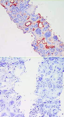
E-Selectin/CD62E in LPS treated Human Kidney Tissue.
E-Selectin/CD62E was detected in acetone fixed cryosections of human kidney tissue using Goat Anti-Human E-Selectin/CD62E Polyclonal Antibody (Catalog # BBA18) overnight at 4 ° C. Tissues were stained with rabbit anti-goat secondary antibody and HRP polymer-conjugated anti-rabbit IgG followed by AEC+Substrate Chromogen (red) followed by counterstaining with hematoxylin (blue). Before incubation with the primary antibody, tissues were treatedex vivowith LPS and also treated to block endogenous peroxidase activity. The lower panel shows an absence of labeling when primary antibody is omitted. Experiments were carried out and images were provided by Dr. Grietje Molema, University of Groningen, The Netherlands.
Detection of E-Selectin/CD62E by Immunohistochemistry
Neutrophil transmigration hotspots are located at ICAM‐1 high expressing cellsSimplified workflow for photoconversion experiments. (1) Neutrophil flow assays were performed in widefield mode with overnight TNF alpha‐treated HUVECs expressing mEos4b. (2) The same field of views were imaged in confocal mode. (3) Areas where hotspots had appeared in the widefield video were converted from green to red using a 405 nm laser. (4) The slides were fixed and the nuclei and an adhesion molecule were stained. The original fields of view were found backing by looking for red signal. (5) In the original video, all successful and unsuccessful adhesion events were annotated. (6) The adhesion spots were overlayed with the stained image. Scale bar, 100 μm.Inverted greyscale LUT of immunofluorescence stains on overnight TNF alpha‐treated HUVECs for ICAM‐1, ICAM‐2, VCAM‐1, and E‐selectin of areas of which time lapses were made, relocated by photoconversion of mEos4b. With yellow asterisks, adhesion events that led to diapedesis are shown. With magenta asterisks, adhesion events that were followed by detachment are shown. Scale bar, 100 μm.Quantification of adhesion events on overnight TNF alpha‐treated HUVEC cells ranked by their preference for diapedesis for expression levels of ICAM‐1 (green dots, 254 diapedesis adhesion events and 57 detachment adhesion events), ICAM‐2 (magenta dots 129 diapedesis adhesion events and 79 detachment adhesion events) VCAM‐1 (blue dots 163 diapedesis adhesion events and 72 detachment adhesion events) and E‐selectin (dark yellow dots = 199 diapedesis adhesion events and 78 detachment adhesion events). Data shown are from 9 (ICAM‐1 and VCAM‐1) or 6 (ICAM‐2 and E‐selectin) images from 3 biological replicates. Median with 95% CI is shown. Image collected and cropped by CiteAb from the following open publication (https://pubmed.ncbi.nlm.nih.gov/36382783), licensed under a CC-BY license. Not internally tested by R&D Systems.Applications for Human E-Selectin/CD62E Antibody
Application
Recommended Usage
Immunohistochemistry
1:100 dilution
Sample: Immersion fixed paraffin-embedded sections of human kidney cancer tissue subjected to Antigen Retrieval Reagent-Basic (Catalog # CTS013) and acetone fixed frozen sections of human normal kidney tissue
Sample: Immersion fixed paraffin-embedded sections of human kidney cancer tissue subjected to Antigen Retrieval Reagent-Basic (Catalog # CTS013) and acetone fixed frozen sections of human normal kidney tissue
Western Blot
1:1000 dilution
Sample: Recombinant Human E-Selectin/CD62E Fc Chimera (Catalog # 724-ES)
Sample: Recombinant Human E-Selectin/CD62E Fc Chimera (Catalog # 724-ES)
Reviewed Applications
Read 2 reviews rated 4.5 using BBA18 in the following applications:
Formulation, Preparation, and Storage
Reconstitution
Reconstitute in 0.5 mL of sterile water.
Formulation
Lyophilized from a 0.2 μm filtered solution in Serum.
Shipping
The product is shipped at ambient temperature. Upon receipt, store it immediately at the temperature recommended below.
Stability & Storage
Use a manual defrost freezer and avoid repeated freeze-thaw cycles.
- 12 months from date of receipt, -20 to -70 °C as supplied.
- 1 month, 2 to 8 °C under sterile conditions after reconstitution.
- 6 months, -20 to -70 °C under sterile conditions after reconstitution.
Background: E-Selectin/CD62E
E-Selectin (Endothelial Leukocyte Adhesion Molecule-1, ELAM-1, CD62E), a member of the Selectin family, is a 107 - 115 kDa cell surface glycoprotein. It is transiently expressed on vascular endothelial cells in response to IL-1 beta and TNF-alpha, and demonstrates peak expression at 4 hours, and decay at 24 hours, in response to activation. E-Selectin ligands, expressed on neutrophils, monocytes, and a subset of memory T cells, are sialylated, fucosylated molecules which bind to the lectin domain of E-Selectin. Immunocytochemical techniques have demonstrated the expression of E-Selectin on healthy and diseased tissue. The human and mouse
E‑Selectin proteins share 81% amino acid similarity.
E-Selectin mediates the attachment of flowing leukocytes to the blood vessel wall during inflammation by binding to E-Selectin ligands on leukocytes. These interactions are labile and permit leukocytes to roll along the vascular endothelium in the direction of blood flow. This initial interaction is followed by a stronger interaction involving ICAM-1 and VCAM-1 that leads eventually to extravasation of the white blood cell through the blood vessel wall into the extracellular matrix tissue.
ELISA techniques have shown that detectable levels of soluble E-Selectin are present in the biological fluids of apparently normal individuals. Furthermore, a number of studies have reported that levels of E-Selectin may be elevated in subjects with a variety of pathological conditions.
Alternate Names
CD62E, ELAM1, LECAM2, SELE
Gene Symbol
SELE
Additional E-Selectin/CD62E Products
Product Documents for Human E-Selectin/CD62E Antibody
Product Specific Notices for Human E-Selectin/CD62E Antibody
For research use only
Loading...
Loading...
Loading...
Loading...
Loading...