Human CD3 Antibody
R&D Systems, part of Bio-Techne | Catalog # MAB100R
Recombinant Monoclonal Antibody.

Conjugate
Catalog #
Key Product Details
Species Reactivity
Human
Applications
Western Blot, Flow Cytometry, Immunocytochemistry, T Cell Stimulation
Label
Unconjugated
Antibody Source
Recombinant Monoclonal Mouse IgG1 Clone # UCHT1R
Product Specifications
Immunogen
Human thymocytes
Specificity
Recognizes the epsilon-chain of the CD3/T cell antigen receptor complex (McMichael, A.J. et al. (1987) Leucocyte Typing III: White Cell Differentiation Antigens, Oxford University Press, New York; Knapp, W. et al. (1989) Leucocyte Typing IV: White Cell Differentiation Antigens, Oxford University Press, New York; Schlossman, S. et al. (1995) Leucocyte Typing V: White Cell Differentiation Antigens, Oxford University Press, New York).
Clonality
Monoclonal
Host
Mouse
Isotype
IgG1
Endotoxin Level
<0.10 EU per 1 μg of the antibody by the LAL method.
Scientific Data Images for Human CD3 Antibody
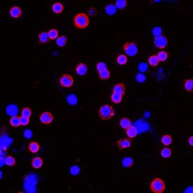
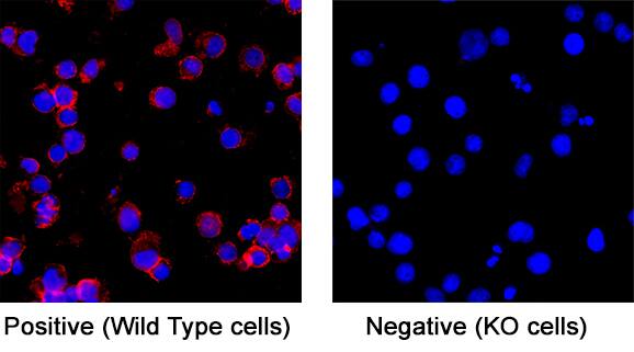
CD3 Specificity is Shown by Immunocytochemistry in Knockout Cell Line.
CD3 was detected in immersion fixed Jurkat human acute T cell leukemia cell line (Positive) and absent in Jurkat CD3 Knockout (Negative) control using Mouse Anti-Human CD3 Monoclonal Antibody (Catalog # MAB100R) at 3 µg/ml for 3 hours at room temperature. Cells were stained using the NorthernLights™ 557-conjugated Anti-Mouse IgG Secondary Antibody (red; Catalog # NL007) and counterstained with DAPI (blue). Specific staining was localized to the cell membrane of wildtype Jurkat cells. View our protocol for Fluorescent ICC Staining of Cells on Coverslips.Detection of Human CD3 epsilon by Western Blot.
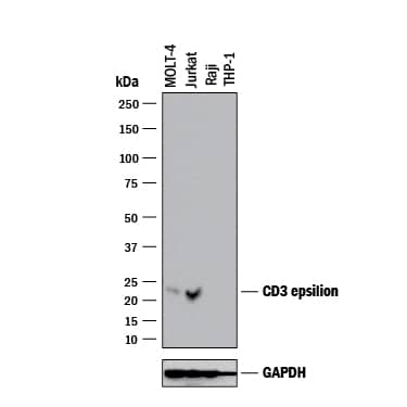
Western blot shows lysates of MOLT-4 human acute lymphoblastic leukemia cell line, Jurkat human acute T cell leukemia cell line, Raji human Burkitt's lymphoma cell line (negative control), and THP-1 human acute monocytic leukemia cell line (negative control). PVDF membrane was probed with 2 µg/mL of Mouse Anti-Human CD3 epsilon Monoclonal Antibody (Catalog # MAB100R) followed by HRP-conjugated Anti-Mouse IgG Secondary Antibody (HAF018). A specific band was detected for CD3 epsilon at approximately 21 kDa (as indicated). GAPDH (Catalog # MAB5718) is shown as a loading control. This experiment was conducted under reducing conditions and using Western Blot Buffer Group 1.Detection of CD3 epsilon in Human Lymphocytes by Flow Cytometry.
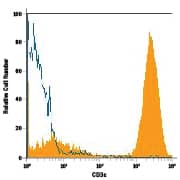
Human peripheral blood lymphocytes were stained with Mouse Anti-Human CD3e Recombinant Monoclonal Antibody (Catalog # MAB100, filled histogram) or isotype control antibody (MAB002, open histogram), followed by Phycoerythrin-conjugated Anti-Mouse IgG Secondary Antibody (F0102B).Applications for Human CD3 Antibody
Application
Recommended Usage
Flow Cytometry
0.25 µg/106 cells
Sample: Human peripheral blood lymphocytes
Sample: Human peripheral blood lymphocytes
Immunocytochemistry
1-25 µg/mL
Sample: Immersion fixed human peripheral blood mononuclear cells (PBMCs) and Jurkat human acute T cell leukemia cell line
Sample: Immersion fixed human peripheral blood mononuclear cells (PBMCs) and Jurkat human acute T cell leukemia cell line
T Cell Stimulation
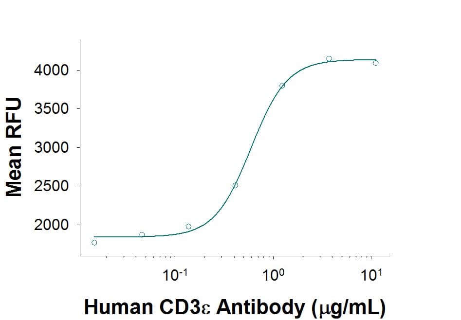
This antibody can be used to activate T cells when immobilized at 1-10 μg/mL (100 μL/well).
Western Blot
2 µg/mL
Sample: MOLT‑4 human acute lymphoblastic leukemia cell line and Jurkat human acute T cell leukemia cell line
Sample: MOLT‑4 human acute lymphoblastic leukemia cell line and Jurkat human acute T cell leukemia cell line
Reviewed Applications
Read 2 reviews rated 5 using MAB100R in the following applications:
Formulation, Preparation, and Storage
Purification
Protein A or G purified from cell culture supernatant
Reconstitution
Reconstitute at 0.5 mg/mL in sterile PBS. For liquid material, refer to CoA for concentration.
Formulation
Lyophilized from a 0.2 μm filtered solution in PBS with Trehalose. See Certificate of Analysis for details.
*Small pack size (-SP) is supplied either lyophilized or as a 0.2 µm filtered solution in PBS.
*Small pack size (-SP) is supplied either lyophilized or as a 0.2 µm filtered solution in PBS.
Shipping
Lyophilized product is shipped at ambient temperature. Liquid small pack size (-SP) is shipped with polar packs. Upon receipt, store immediately at the temperature recommended below.
Stability & Storage
Use a manual defrost freezer and avoid repeated freeze-thaw cycles.
- 12 months from date of receipt, -20 to -70 °C as supplied.
- 1 month, 2 to 8 °C under sterile conditions after reconstitution.
- 6 months, -20 to -70 °C under sterile conditions after reconstitution.
Background: CD3
CD3 epsilon is one of at least four invariant proteins that associate with the variable antigen recognition chains of the T cell receptor and function in signal transduction.
References
- Beverly, P.C.L. and R.E. Callard (1981) Eur. J. Immunol. 11:329.
Alternate Names
CD_antigen: CD3e, CD3 antigen, delta subunit, CD3d antigen, CD3d antigen, delta polypeptide (TiT3 complex), CD3d molecule, delta (CD3-TCR complex), CD3-DELTA, CD3e, CD3e antigen, CD3e antigen, epsilon polypeptide (TiT3 complex), CD3e molecule, epsilon (CD3-TCR complex), CD3-epsilon, CD3G, CD3g antigen, CD3g antigen, gamma polypeptide (TiT3 complex), CD3g molecule, epsilon (CD3-TCR complex), CD3g molecule, gamma (CD3-TCR complex), CD3-GAMMA, FLJ17620, FLJ17664, FLJ18683, FLJ79544, FLJ94613, IMD18, MGC138597, T3DOKT3, delta chain, T3E, T-cell antigen receptor complex, epsilon subunit of T3, T-cell receptor T3 delta chain, T-cell surface antigen T3/Leu-4 epsilon chain, T-cell surface glycoprotein CD3 delta chain, T-cell surface glycoprotein CD3 epsilon chain, TCRE
Gene Symbol
CD3E
Additional CD3 Products
Product Documents for Human CD3 Antibody
Product Specific Notices for Human CD3 Antibody
For research use only
Loading...
Loading...
Loading...
Loading...
Loading...
