
ATF4 Antibody (SD20-92)
Novus Biologicals, part of Bio-Techne | Catalog # NBP2-67766
Recombinant Monoclonal Antibody

![Immunohistochemistry-Paraffin: ATF4 Antibody (SD20-92) [NBP2-67766]](https://resources.bio-techne.com/images/products/ATF4-Antibody-SD20-92-Immunohistochemistry-Paraffin-NBP2-67766-img0017.jpg "Immunohistochemistry-Paraffin: ATF4 Antibody (SD20-92) [NBP2-67766]")
Scientific images may be resized, cropped, or adjusted for brightness or contrast for presentation purposes. These updates are limited to presentation and do not affect the underlying scientific interpretation of the data. Where available, additional source data may be provided upon request.
Key Product Details
Validated by
Biological Validation
Species Reactivity
Validated:
Human, Mouse, Rat
Cited:
Mouse
Applications
Validated:
Immunohistochemistry, Immunohistochemistry-Paraffin, Western Blot, Flow Cytometry, Immunocytochemistry/ Immunofluorescence, Immunoprecipitation
Cited:
Immunohistochemistry-Frozen, IF/IHC
Label
Unconjugated
Antibody Source
Recombinant Monoclonal Rabbit IgG Clone # SD20-92 expressed in HEK293
Concentration
1 mg/ml
Product Specifications
Immunogen
Recombinant protein within Human ATF4 aa 1-220 / 351. (SwissProt: P18848 Human; SwissProt: Q06507 Mouse; SwissProt: Q9ES19 Rat)
Localization
Nucleus, Nucleus speckle, Cell membrane, Cytoplasm, Centrosome.
Clonality
Monoclonal
Host
Rabbit
Isotype
IgG
Scientific Data Images for ATF4 Antibody (SD20-92)
Detection of Human ATF4 by Simple Western TM.
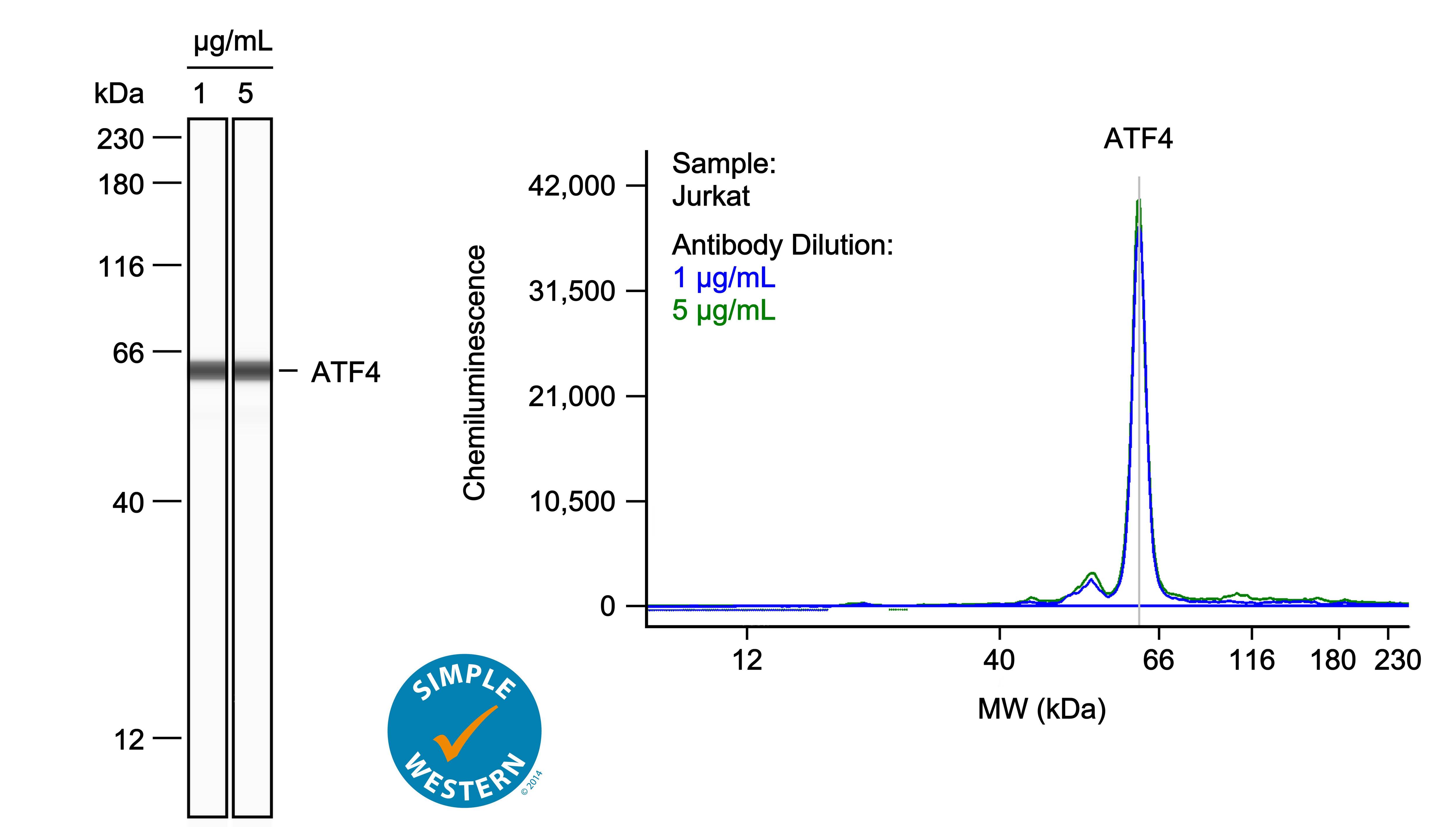
Left: Simple Western lane view shows lysates of Jurkat human acute T cell leukemia cell line, loaded at 1 mg/ml. A specific band was detected for ATF4 at approximately 63 kDa (as indicated) using both 1 ug/ml and 5 ug/ml of Rabbit Anti-Human ATF4 Monoclonal Antibody (Catalog # NBP2-67766) followed by HRP-conjugated Goat Anti-Rabbit Secondary Antibody (Catalog # 042-206). This experiment was conducted under reducing conditions and using the 12-230kDa separation system. Right: Simple Western electropherogram showing the same Rabbit Anti-Human ATF4 Monoclonal Antibody (Catalog # NBP2-67766) tested at 1 ug/ml (blue line) and 5 ug/ml (green line) in the Jurkat human acute T cell leukemia cell line.![Western Blot: ATF4 Antibody (SD20-92) [NBP2-67766]](https://resources.bio-techne.com/images/products/ATF4-Antibody-SD20-92-Western-Blot-NBP2-67766-img0007.jpg "Western Blot: ATF4 Antibody (SD20-92) [NBP2-67766]")
Western Blot: ATF4 Antibody (SD20-92) [NBP2-67766]
Western Blot: ATF4 Antibody (SD20-92) [NBP2-67766] - Analysis of ATF4 on different lysates. Proteins were transferred to a PVDF membrane and blocked with 5% BSA in PBS for 1 hour at room temperature. The primary antibody (1:500) was used in 5% BSA at room temperature for 2 hours. Goat Anti-Rabbit IgG - HRP Secondary Antibody at 1:5,000 dilution was used for 1 hour at room temperature. Positive control: Lane 1: HeLa cell lysateLane 2: PC-12 cell lysate Lane 3: HL-60 cell lysate Lane 4: K562 cell lysateLane 5: human lung carcinoma tissue lysate.![Immunocytochemistry/ Immunofluorescence: ATF4 Antibody (SD20-92) [NBP2-67766]](https://resources.bio-techne.com/images/products/ATF4-Antibody-SD20-92-Immunocytochemistry-Immunofluorescence-NBP2-67766-img0005.jpg "Immunocytochemistry/ Immunofluorescence: ATF4 Antibody (SD20-92) [NBP2-67766]")
Immunocytochemistry/ Immunofluorescence: ATF4 Antibody (SD20-92) [NBP2-67766]
Immunocytochemistry/Immunofluorescence: ATF4 Antibody (SD20-92) [NBP2-67766] - Staining ATF4 in N2A cells (green). The nuclear counter stain is DAPI (blue). Cells were fixed in paraformaldehyde, permeabilised with 0.25% Triton X-100/PBS.![Flow Cytometry: ATF4 Antibody (SD20-92) [NBP2-67766]](https://resources.bio-techne.com/images/products/ATF4-Antibody-SD20-92-Flow-Cytometry-NBP2-67766-img0001.jpg "Flow Cytometry: ATF4 Antibody (SD20-92) [NBP2-67766]")
![Immunohistochemistry-Paraffin: ATF4 Antibody (SD20-92) [NBP2-67766]](https://resources.bio-techne.com/images/products/ATF4-Antibody-SD20-92-Immunohistochemistry-Paraffin-NBP2-67766-img0008.jpg "Immunohistochemistry-Paraffin: ATF4 Antibody (SD20-92) [NBP2-67766]")
![Immunohistochemistry-Paraffin: ATF4 Antibody (SD20-92) [NBP2-67766]](https://resources.bio-techne.com/images/products/ATF4-Antibody-SD20-92-Immunohistochemistry-Paraffin-NBP2-67766-img0009.jpg "Immunohistochemistry-Paraffin: ATF4 Antibody (SD20-92) [NBP2-67766]")
![Immunohistochemistry-Paraffin: ATF4 Antibody (SD20-92) [NBP2-67766]](https://resources.bio-techne.com/images/products/ATF4-Antibody-SD20-92-Immunohistochemistry-Paraffin-NBP2-67766-img0010.jpg "Immunohistochemistry-Paraffin: ATF4 Antibody (SD20-92) [NBP2-67766]")
![Immunohistochemistry-Paraffin: ATF4 Antibody (SD20-92) [NBP2-67766]](https://resources.bio-techne.com/images/products/ATF4-Antibody-SD20-92-Immunohistochemistry-Paraffin-NBP2-67766-img0011.jpg "Immunohistochemistry-Paraffin: ATF4 Antibody (SD20-92) [NBP2-67766]")
![Immunohistochemistry-Paraffin: ATF4 Antibody (SD20-92) [NBP2-67766]](https://resources.bio-techne.com/images/products/ATF4-Antibody-SD20-92-Immunohistochemistry-Paraffin-NBP2-67766-img0012.jpg "Immunohistochemistry-Paraffin: ATF4 Antibody (SD20-92) [NBP2-67766]")
![Immunohistochemistry-Paraffin: ATF4 Antibody (SD20-92) [NBP2-67766]](https://resources.bio-techne.com/images/products/ATF4-Antibody-SD20-92-Immunohistochemistry-Paraffin-NBP2-67766-img0014.jpg "Immunohistochemistry-Paraffin: ATF4 Antibody (SD20-92) [NBP2-67766]")
![Immunohistochemistry-Paraffin: ATF4 Antibody (SD20-92) [NBP2-67766]](https://resources.bio-techne.com/images/products/ATF4-Antibody-SD20-92-Immunohistochemistry-Paraffin-NBP2-67766-img0015.jpg "Immunohistochemistry-Paraffin: ATF4 Antibody (SD20-92) [NBP2-67766]")
![Immunohistochemistry-Paraffin: ATF4 Antibody (SD20-92) [NBP2-67766]](https://resources.bio-techne.com/images/products/ATF4-Antibody-SD20-92-Immunohistochemistry-Paraffin-NBP2-67766-img0016.jpg "Immunohistochemistry-Paraffin: ATF4 Antibody (SD20-92) [NBP2-67766]")
Applications for ATF4 Antibody (SD20-92)
Application
Recommended Usage
Flow Cytometry
1:50-1:100
Immunocytochemistry/ Immunofluorescence
1:50-1:200
Immunohistochemistry-Paraffin
1:50-1:200
Western Blot
1:500-1:2000
Reviewed Applications
Read 1 review rated 1 using NBP2-67766 in the following applications:
Formulation, Preparation, and Storage
Purification
Protein A purified
Formulation
TBS (pH7.4), 0.05% BSA, 40% Glycerol
Preservative
0.05% Sodium Azide
Concentration
1 mg/ml
Shipping
The product is shipped with polar packs. Upon receipt, store it immediately at the temperature recommended below.
Stability & Storage
Store at 4C short term. Aliquot and store at -20C long term. Avoid freeze-thaw cycles.
Background: ATF4
Long Name
Activating Transcription Factor 4
Alternate Names
TAXREB67, TXREB
Gene Symbol
ATF4
Additional ATF4 Products
Product Documents for ATF4 Antibody (SD20-92)
Product Specific Notices for ATF4 Antibody (SD20-92)
This product is for research use only and is not approved for use in humans or in clinical diagnosis. Primary Antibodies are guaranteed for 1 year from date of receipt.
Loading...
Loading...
Loading...
Loading...
Loading...