

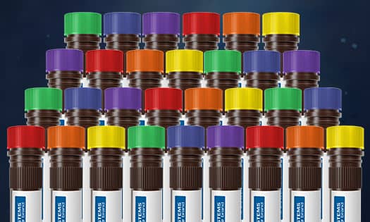
Increased Flexibility in Flow Panel Design
Increased Flexibility in Flow Panel Design
Explore an expansive catalog of mFluor™ Violet Conjugated Antibodies for enhanced versatility in your experiments.
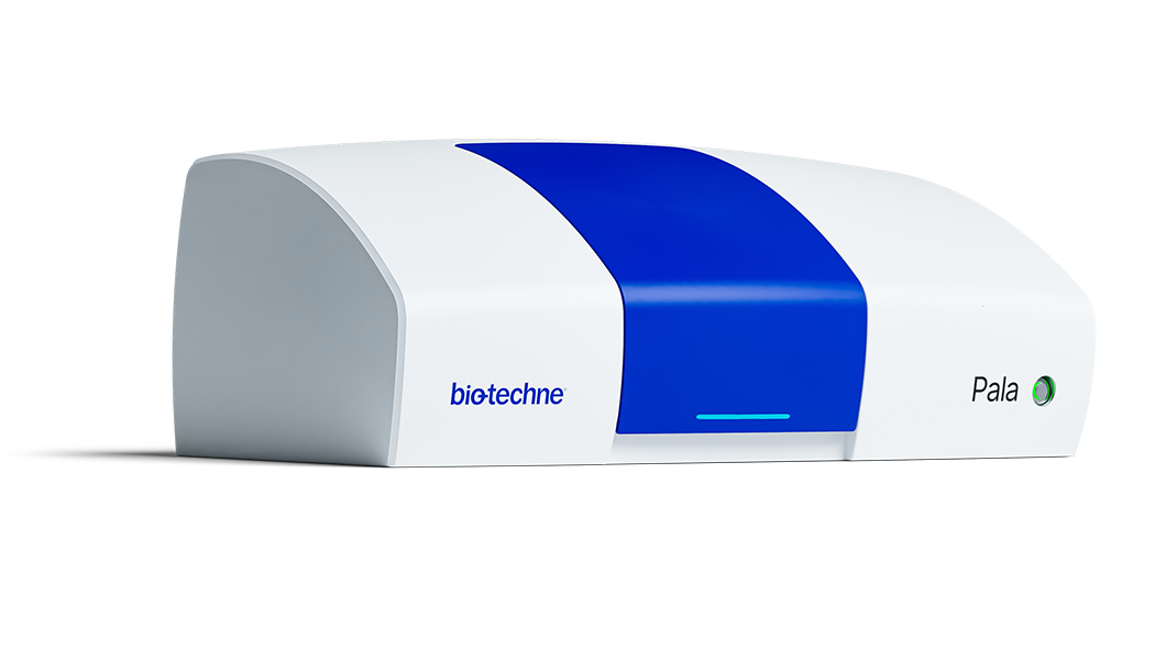
Fast, Gentle & Easy Single Cell Isolation
Fast, Gentle & Easy Single Cell Isolation
The Pala Cell Sorter and Single Cell Dispenser makes single cell sorting fast and easy. See how this benchtop instrument combine microfluidics, flow cytometry and direct liquid dispensing to accomplish gentle cell sorting and dispensing in one simple step.

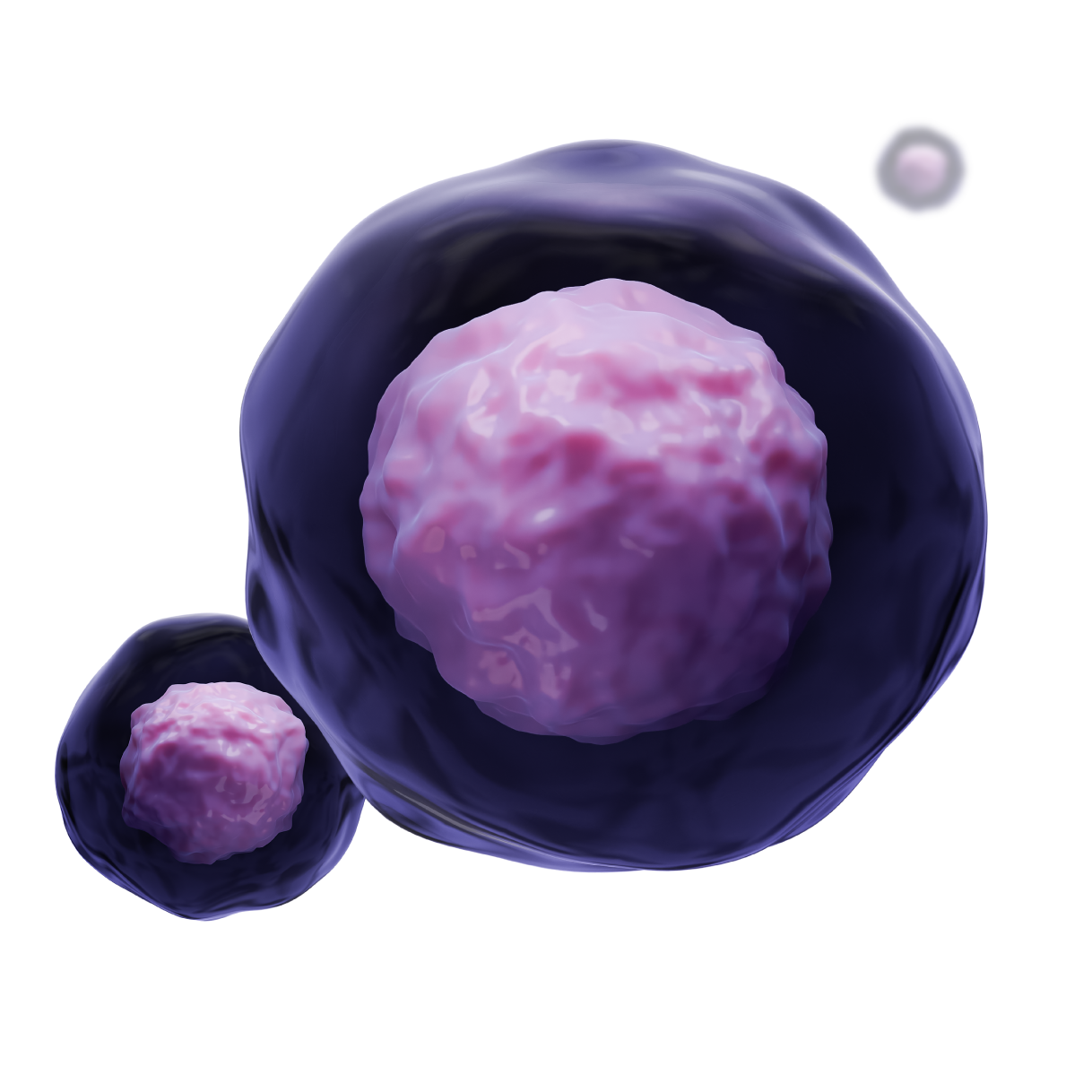
Fluorokines™: Fluorescently Labeled Proteins
Fluorokines™: Fluorescently Labeled Proteins
Easily measure transduction efficiency of CAR T or CAR NK cell preparations or determine B cell specificity after COVID-19 infection or vaccination.
Find our full selection of flow cytometry validated antibodies, including fluorescent conjugated primary antibodies, secondary antibodies and isotype controls. Explore our broad selection of fluorescently conjugated antibodies, including Alexa Fluor® 488, Alexa Fluor® 647, PE, PE-Cy7™, and more! Take the guesswork out of flow cytometry panel design with our multicolor flow kits and panels.
- Cell Surface Staining Protocol (at R&D Systems)
- Intracellular Staining Protocol (Alcohols) (at R&D Systems)
- Intracellular Staining Protocol (Detergents) (at R&D Systems)
- Flow Cytometry Trouble Shooting Guide

Flow Cytometry Panel Builder Tool
Flow Cytometry Panel Builder Tool
Save time and reduce costly mistakes by quickly finding compatible reagents using the Panel Builder Tool.

Interactive Cell Marker Tool
Interactive Cell Marker Tool
Learn more about cell surface and intracellular markers from R&D Systems, a Bio‑Techne brand.
Meet MitoBrilliant™ Dyes

MagCellect™ Cell Selection Kits & Reagents
Isolate and enrich cell populations through magnetic, bead-based positive or negative selection without the need for specialized columns.
Featured MagCellect Kits

Flow Cytometry Handbook
Our Flow Cytometry Handbook is the must have resource for flow cytometry researchers. Download now to find step-by-step protocols and troubleshooting tips.
Flow cytometry is a high throughput, single-cell analysis technique that uses lasers to rapidly activate fluorescently-labeled conjugated antibodies on cells in a suspended solution. It is a powerful tool as it can measure many parameters on single cells, as well as very quickly collect information from millions of cells. In flow cytometry, single-cell suspensions are passed through single or multiple lasers, which causes the light to scatter. Cell populations can be sorted based on light scatter, where forward scatter provides cell size analysis and side scatter determines cell granularity. Flow cytometry can provide information about cell size and volume, evaluate the purity of isolated cell subpopulations, characterize different cell types in a heterogeneous population, while providing a technique that can examine the expression of both cell surface and intracellular proteins.
Fluorophore compounds, such as fluorescently-labeled antibodies or propidium iodide, are commonly used in flow cytometry to detect proteins of interest. Using fluorophores with nonoverlapping maximum emission spectra enables simultaneous individual detection of fluorescently stained cells. Isotype control antibodies are used as negative controls to validate a flow cytometry experiment. Multiple research areas including cancer biology, immunology, infectious disease monitoring, virology, and molecular biology utilize flow cytometry.
What is FACS?
Fluorescence-activated cell sorting (FACS) is a specialized type of flow cytometry. It provides a method for sorting a heterogeneous mixture of biological cells into separate containers, one cell at a time. Cell sorting is based upon the specific light scattering and fluorescent characteristics of each cell.
What is the minimum number of cells needed for flow cytometry?
The number of cells needed for a particular experiment is dependent on your needs. You would need fewer cells if you are probing for a robustly expressed antigen compared to a sparsely expressed protein. Additionally, you would need more cells for multicolor flow cytometry versus single color staining. However, we recommend starting with 1 × 10⁶ cells per 100 µl of staining buffer per tube.
What controls are used in flow cytometry?
Flow cytometry controls can be split into the following categories.
- Instrument Controls: Calibration particles or fluorescent beads from the instrument’s manufacturer should be run prior to use to ensure the instrument is performing properly.
- Compensation Controls: When performing multicolor flow cytometry, the emission spectra from the different fluorophores used can overlap and can spread into multiple channels. The process of correcting for spillover is known as compensation. For compensation, a single stained sample is needed from each fluorochrome in the experiment. These can be set up using cells, compensation beads, or antibody capture beads.
- Gating Controls: One of the most critical aspects of analyzing flow cytometry data is properly setting gates. The Fluorescence Minus One (FMO) control is necessary for interpreting flow data and for setting gates. Since FMO controls provide a reliable assessment of background in a multicolor experiment, they can accurately discriminate between negative and positive cell populations.
- Cell Viability Controls: Dead cells can produce false positives because they autofluoresce and display an increased affinity for non-specific antibody binding. As a result, these dead cells need to be removed from data analysis. Several fluorescent dyes, such as 7-AAD, calcein AM, and propidium iodide, can be used to distinguish dead cells from live cells.
- Unstained Cells Controls: Several cell components, such as flavin coenzymes and NADPH, are endogenous fluorophores that exhibit autofluorescence. Running a sample of unstained cells through the flow cytometry will allow you to determine the amount of background fluorescence due to autofluorescence so that you can gate appropriately.
- Isotype Controls: Isotype controls are used to assess non-specific binding due to antibody class. An isotype control is an antibody that lacks specificity to the target of interest, but matches the class, type, and fluorescent conjugate of the primary antibody.
Products for Differentiating and Expanding Cells
Resources