
MyoD Antibody (5.8A) [FITC]
Novus Biologicals, part of Bio-Techne | Catalog # NB100-56511F

Conjugate
Catalog #
Forumulation
Catalog #
Key Product Details
Species Reactivity
Validated:
Human, Mouse
Predicted:
Porcine (100%), Rat (100%). Backed by our 100% Guarantee.
Applications
Knockout Validated, Immunohistochemistry, Immunohistochemistry-Paraffin, Immunohistochemistry-Frozen, Western Blot, Flow Cytometry, Immunocytochemistry/ Immunofluorescence, Immunoprecipitation, Knockdown Validated
Label
FITC (Excitation = 495 nm, Emission = 519 nm)
Antibody Source
Monoclonal Mouse IgG1 kappa Clone # 5.8A
Concentration
Please see the vial label for concentration. If unlisted please contact technical services.
Product Specifications
Immunogen
The 5.8A antibody was made against recombinant mouse MyoD protein but it also recognizes human (myf3), rat, and cat homologs. The epitope of this antibody was mapped to a region within aa 180-189 of mouse MyoD (NP_002469).
Specificity
In Rh-30, a ~45 kDa band should be observed.
Clonality
Monoclonal
Host
Mouse
Isotype
IgG1 kappa
Description
This conjugate is made on demand and is not held in inventory. Each order represents a unique lot. All conjugates are generated using validated conjugation protocols and meet strict degree of labeling (F/P) specifications; additional information is available upon request. The antibody concentration is provided on the individual product label.
For specialized conjugation needs, like large quantities, specific concentrations, and defined F/P ratios, bulk and custom antibody conjugation services are available.
For specialized conjugation needs, like large quantities, specific concentrations, and defined F/P ratios, bulk and custom antibody conjugation services are available.
Scientific Data Images for MyoD Antibody (5.8A) [FITC]
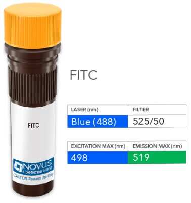
Product Image: MyoD Antibody (5.8A) [FITC] [NB100-56511F] - Vial of FITC conjugated antibody. FITC is optimally excited at 498 nm by the Blue laser (488 nm) and has an emission maximum of 519 nm.
![MyoD Antibody (5.8A) [FITC]](https://resources.bio-techne.com/images/products/nb100-56511f_mouse-monoclonal-myod-antibody-5-8a-fitc-flow-cytometry-31122024154538..jpg "Detection of MyoD (5.8A) in SJCRH30 Human Cell Line by Flow Cytometry.")
Detection of MyoD (5.8A) in SJCRH30 Human Cell Line by Flow Cytometry.
An intracellular stain was performed on SJCRH30 human Rhabdomyosarcoma cell line with Mouse anti- MyoD (5.8A) Protein-G purified Monoclonal Antibody conjugated to FITC (Catalog # NB100-56511F, blue histogram) or matched control antibody (orange histogram) at 5 µg/mL for 30 minutes at RT.Applications for MyoD Antibody (5.8A) [FITC]
Application
Recommended Usage
Flow Cytometry
Optimal dilutions of this antibody should be experimentally determined.
Immunocytochemistry/ Immunofluorescence
Optimal dilutions of this antibody should be experimentally determined.
Immunohistochemistry
Optimal dilutions of this antibody should be experimentally determined.
Immunohistochemistry-Frozen
Optimal dilutions of this antibody should be experimentally determined.
Immunohistochemistry-Paraffin
Optimal dilutions of this antibody should be experimentally determined.
Immunoprecipitation
Optimal dilutions of this antibody should be experimentally determined.
Knockdown Validated
Optimal dilutions of this antibody should be experimentally determined.
Knockout Validated
Optimal dilutions of this antibody should be experimentally determined.
Western Blot
Optimal dilutions of this antibody should be experimentally determined.
Application Notes
Recommended applications are based on validated applications from the unconjugated base product NB100-56511. This conjugated antibody is not kept in inventory and is made to order using validated conjugation protocols.
Spectra Viewer
Plan Your Experiments
Use our spectra viewer to interactively plan your experiments, assessing multiplexing options. View the excitation and emission spectra for our fluorescent dye range and other commonly used dyes.
Spectra Viewer
Formulation, Preparation, and Storage
Purification
Protein G purified
Formulation
PBS
Preservative
0.05% Sodium Azide
Concentration
Please see the vial label for concentration. If unlisted please contact technical services.
Shipping
The product is shipped with polar packs. Upon receipt, store it immediately at the temperature recommended below.
Stability & Storage
Store at 4C in the dark.
Background: MyoD
Long Name
Myoblast Determination Protein 1
Alternate Names
bHLHc1, MYF3, MYOD1, PUM
Gene Symbol
MYOD1
Additional MyoD Products
Product Documents for MyoD Antibody (5.8A) [FITC]
Product Specific Notices for MyoD Antibody (5.8A) [FITC]
This conjugate is made on demand. Actual recovery may vary from the stated volume of this product. The volume will be greater than or equal to the unit size stated on the datasheet.
This product is for research use only and is not approved for use in humans or in clinical diagnosis. Primary Antibodies are guaranteed for 1 year from date of receipt.
Loading...
Loading...
Loading...
Loading...
Loading...