
Ki67/MKI67 Antibody [DyLight 650]
Novus Biologicals, part of Bio-Techne | Catalog # NB110-89717C

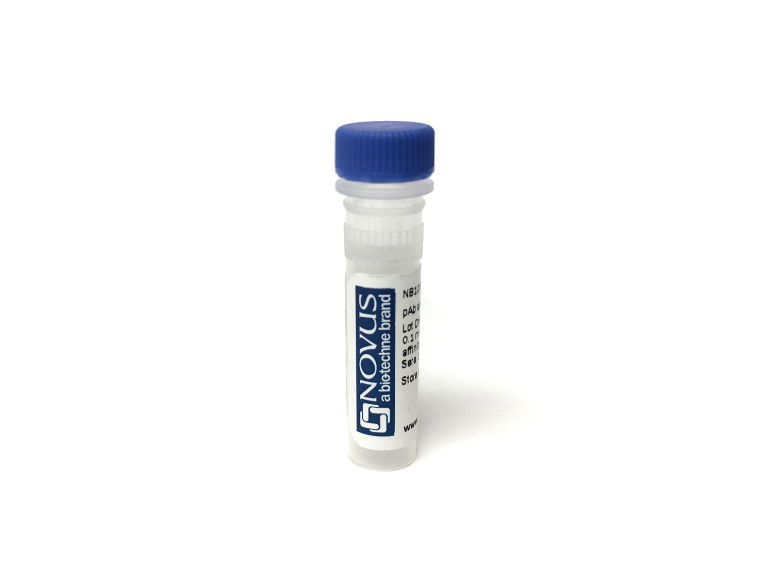
Conjugate
Catalog #
Forumulation
Catalog #
Key Product Details
Species Reactivity
Human, Mouse, Rat, Porcine
Applications
Knockout Validated, Immunohistochemistry, Immunohistochemistry-Paraffin, Immunohistochemistry-Frozen, Western Blot, Flow Cytometry, Immunocytochemistry/ Immunofluorescence
Label
DyLight 650 (Excitation = 652 nm, Emission = 672 nm)
Antibody Source
Polyclonal Rabbit IgG
Concentration
Please see the vial label for concentration. If unlisted please contact technical services.
Product Specifications
Immunogen
The immunogen for this KI67/MKI67 Antibody was made using a synthetic peptide from the internal region of Mouse KI67/MKI67, between aminoacids 1850-1950 (1899-1916) Uniprot# E9PVX6.
Reactivity Notes
Ki67/MKI67 Antibody reacted with Rat in in scientific literature (PMID: 24275061). Use in Porcine reported in scientific literature (PMID:32132871).
Localization
Nuclear
Marker
Proliferation Marker
Clonality
Polyclonal
Host
Rabbit
Isotype
IgG
Description
This conjugate is made on demand and is not held in inventory. Each order represents a unique lot. All conjugates are generated using validated conjugation protocols and meet strict degree of labeling (F/P) specifications; additional information is available upon request. The antibody concentration is provided on the individual product label.
For specialized conjugation needs, like large quantities, specific concentrations, and defined F/P ratios, bulk and custom antibody conjugation services are available.
For specialized conjugation needs, like large quantities, specific concentrations, and defined F/P ratios, bulk and custom antibody conjugation services are available.
Applications for Ki67/MKI67 Antibody [DyLight 650]
Application
Recommended Usage
Flow Cytometry
Optimal dilutions of this antibody should be experimentally determined.
Immunocytochemistry/ Immunofluorescence
Optimal dilutions of this antibody should be experimentally determined.
Immunohistochemistry
Optimal dilutions of this antibody should be experimentally determined.
Immunohistochemistry-Frozen
Optimal dilutions of this antibody should be experimentally determined.
Immunohistochemistry-Paraffin
Optimal dilutions of this antibody should be experimentally determined.
Knockout Validated
Optimal dilutions of this antibody should be experimentally determined.
Western Blot
Optimal dilutions of this antibody should be experimentally determined.
Application Notes
Recommended applications are based on validated applications from the unconjugated base product NB110-89717. This conjugated antibody is not kept in inventory and is made to order using validated conjugation protocols.
Spectra Viewer
Plan Your Experiments
Use our spectra viewer to interactively plan your experiments, assessing multiplexing options. View the excitation and emission spectra for our fluorescent dye range and other commonly used dyes.
Spectra Viewer
Formulation, Preparation, and Storage
Purification
Immunogen affinity purified
Formulation
50mM Sodium Borate
Preservative
0.05% Sodium Azide
Concentration
Please see the vial label for concentration. If unlisted please contact technical services.
Shipping
The product is shipped with polar packs. Upon receipt, store it immediately at the temperature recommended below.
Stability & Storage
Store at 4C in the dark.
Background: Ki67/MKI67
Detection of Ki67 by immunostaining is commonly used as a proliferation marker in solid tumors, as well as certain hematological malignancies (3-5). The Ki67 index, which reports on positive Ki67 stained tumor cell nuclei, has been extensively studied as a prognostic biomarker in cancers such as breast cancer and cervical cancer.
References
1. Gerdes J, Schwab U, Lemke H, Stein H. (1983) Production of a mouse monoclonal antibody reactive with a human nuclear antigen associated with cell proliferation. Int J Cancer. 31:13-20. PMID: 6339421
2. Starborg M, Gell K, Brundell E and Hoog C. (1996) The murine Ki-67 cell proliferation antigen accumulates in the nucleolar and heterochromatic regions of interphase cells and at the periphery of the mitotic chromosomes in a process essential for cell cycle progression. J Cell Sci. 109:143-153. 1996
3. Karamitopoulou E, Perentes E, Tolnay M, Probst A. (1998) Prognostic significance of MIB-1, p53, and bcl-2 immunoreactivity in meningiomas. Hum Pathol. 29(2):140-5. PMID: 9490273
4. Geyer FC, Rodrigues DN, Weigelt B and Reis-Filho JS. (2012) Molecular classification of estrogen receptor-positive/luminal breast cancers. Adv Anat Pathol. 19(1):39-53. PMID: 22156833
5. Ikenberg H, Bergeron C, Schmidt D, Griesser H, Alameda F, Angeloni C, Bogers J, Dachez R, Denton K, Hariri J, Keller T, von Knebel Doeberitz M, Neumann HH, Puig-Tintore LM, Sideri M, Rehm S, Ridder R; PALMS Study Group. (2013) Screening for cervical cancer precursors with p16/Ki-67 dual-stained cytology: results of the PALMS study. J Natl Cancer Inst. 105(20):1550-7. PMID: 24096620
Long Name
Antigen Identified by Monoclonal Antibody Ki67
Alternate Names
Ki-67, KIA, MIB-1, MKI67, PPP1R105, TSG126
Gene Symbol
MKI67
Additional Ki67/MKI67 Products
Product Documents for Ki67/MKI67 Antibody [DyLight 650]
Product Specific Notices for Ki67/MKI67 Antibody [DyLight 650]
DyLight (R) is a trademark of Thermo Fisher Scientific Inc. and its subsidiaries.
This product is for research use only and is not approved for use in humans or in clinical diagnosis. Primary Antibodies are guaranteed for 1 year from date of receipt.
Loading...
Loading...
Loading...
Loading...
Loading...
Loading...