
Mouse TLR4 Antibody
R&D Systems, part of Bio-Techne | Catalog # MAB27591
Recombinant Monoclonal Antibody.


Scientific images may be resized, cropped, or adjusted for brightness or contrast for presentation purposes. These updates are limited to presentation and do not affect the underlying scientific interpretation of the data. Where available, additional source data may be provided upon request.
Conjugate
Catalog #
Key Product Details
Species Reactivity
Validated:
Mouse
Cited:
Mouse, Transgenic Mouse
Applications
Validated:
Flow Cytometry, Immunocytochemistry
Cited:
Flow Cytometry, Immunocytochemistry, Bioassay
Label
Unconjugated
Antibody Source
Recombinant Monoclonal Rabbit IgG Clone # 1203B
Product Specifications
Immunogen
Mouse myeloma cell line NS0-derived recombinant mouse TLR4
Asn26-Lys629
Accession # Q9QUK6
Asn26-Lys629
Accession # Q9QUK6
Specificity
Detects mouse TLR4 in direct ELISAs.
Clonality
Monoclonal
Host
Rabbit
Isotype
IgG
Scientific Data Images for Mouse TLR4 Antibody
Detection of TLR4 in RAW 264.7 Mouse Cell Line by Flow Cytometry.
RAW 264.7 mouse monocyte/macrophage cell line was stained with Rabbit Anti-Mouse TLR4 Monoclonal Antibody (Catalog # MAB27591, filled histogram) or isotype control antibody (Catalog # AB-105-C, open histogram), followed by Phycoerythrin-conjugated Anti-Rabbit IgG Secondary Antibody (Catalog # F0110).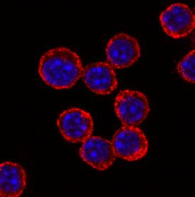
TLR4 in RAW 264.7 Mouse Cell Line.
TLR4 was detected in immersion fixed RAW 264.7 mouse monocyte/macrophage cell line using Rabbit Anti-Mouse TLR4 Monoclonal Antibody (Catalog # MAB27591) at 1 µg/mL for 3 hours at room temperature. Cells were stained using the NorthernLights™ 557-conjugated Anti-Rabbit IgG Secondary Antibody (red; Catalog # NL004) and counterstained with DAPI (blue). Specific staining was localized to cell surfaces and cytoplasm. View our protocol for Fluorescent ICC Staining of Non-adherent Cells.
Detection of TLR4 by Immunocytochemistry/ Immunofluorescence
(s)uPAR is a part of TLR4 interactome. (A) Primary peritoneal macrophages from uPAR−/− mice were stimulated with suPAR with or without LPS for 15 min. Then, cells were fixed and stained for TLR4 (Alexa 488, green) and uPAR (Alexa 647, red). DAPI was used as nuclear stain. Scale bar 10 μm. (B) Primary WT and uPAR−/− macrophages were stimulated with 50 ng/ml LPS and 1 μg/ml suPAR for 3 h. Data are presented as mean ± SD from two independent experiments performed in duplicates with pooled PM isolated from n=4 mice per group. *P < 0.05; **P < 0.01; ***P < 0.001. (C) Raw 264.7 cells were stimulated with 100 ng/ml LPS, fixed and stained as in A. Scale bar 12.5 μm. (D) Raw 264.7 cells were stimulated with 1 μg/ml biotin-LPS for 30 min, then cell lysis was performed. Protein complexes were precipitated using streptavidin magnetic beads and analyzed by western blotting using anti-murine uPAR antibody. One of 3 independent experiments with similar results is shown. Image collected and cropped by CiteAb from the following open publication (https://pubmed.ncbi.nlm.nih.gov/33362762), licensed under a CC-BY license. Not internally tested by R&D Systems.Applications for Mouse TLR4 Antibody
Application
Recommended Usage
Flow Cytometry
0.25 µg/106 cells
Sample: RAW 264.7 mouse monocyte/macrophage cell line
Sample: RAW 264.7 mouse monocyte/macrophage cell line
Immunocytochemistry
1-25 µg/mL
Sample: Immersion fixed RAW 264.7 mouse monocyte/macrophage cell line
Sample: Immersion fixed RAW 264.7 mouse monocyte/macrophage cell line
Formulation, Preparation, and Storage
Purification
Protein A or G purified from cell culture supernatant
Reconstitution
Reconstitute at 0.5 mg/mL in sterile PBS. For liquid material, refer to CoA for concentration.
Formulation
Lyophilized from a 0.2 μm filtered solution in PBS with Trehalose. *Small pack size (SP) is supplied either lyophilized or as a 0.2 µm filtered solution in PBS.
Shipping
Lyophilized product is shipped at ambient temperature. Liquid small pack size (-SP) is shipped with polar packs. Upon receipt, store immediately at the temperature recommended below.
Stability & Storage
Use a manual defrost freezer and avoid repeated freeze-thaw cycles.
- 12 months from date of receipt, -20 to -70 °C as supplied.
- 1 month, 2 to 8 °C under sterile conditions after reconstitution.
- 6 months, -20 to -70 °C under sterile conditions after reconstitution.
Background: TLR4
TLR4 belongs to the IL-1 receptor/Toll-like receptor superfamily. It is expressed in placenta and peripheral blood leukocytes. TLR4 is the pattern recognition receptor for LPS.
Long Name
Toll-like Receptor 4
Alternate Names
CD284
Gene Symbol
TLR4
UniProt
Additional TLR4 Products
Product Documents for Mouse TLR4 Antibody
Product Specific Notices for Mouse TLR4 Antibody
For research use only
Loading...
Loading...
Loading...
Loading...
Loading...
Loading...