
Mouse CCL3/MIP-1 alpha Antibody
R&D Systems, part of Bio-Techne | Catalog # AB-450-NA


Scientific images may be resized, cropped, or adjusted for brightness or contrast for presentation purposes. These updates are limited to presentation and do not affect the underlying scientific interpretation of the data. Where available, additional source data may be provided upon request.
Key Product Details
Validated by
Biological Validation
Species Reactivity
Validated:
Mouse
Cited:
Mouse
Applications
Validated:
Western Blot, Neutralization
Cited:
Western Blot, Neutralization, In vivo assay
Label
Unconjugated
Antibody Source
Polyclonal Goat IgG
Product Specifications
Immunogen
E. coli-derived recombinant mouse CCL3/MIP-1 alpha (R&D Systems, Catalog # 450-MA)
Ala24-Ala92
Accession # Q5QNW0
Ala24-Ala92
Accession # Q5QNW0
Specificity
Detects mouse CCL3/MIP-1 alpha in direct ELISAs and Western blots. In Western blots, less than 5% cross-reactivity with recombinant human (rh) MIP-1 beta, rhMIP-1 alpha, and recombinant mouse MIP-1 beta is observed.
Clonality
Polyclonal
Host
Goat
Isotype
IgG
Endotoxin Level
<0.10 EU per 1 μg of the antibody by the LAL method.
Scientific Data Images for Mouse CCL3/MIP-1 alpha Antibody
Chemotaxis Induced by CCL3/MIP‑1 alpha and Neutrali-zation by Mouse CCL3/MIP‑1 alpha Antibody.
Recombinant Mouse CCL3/MIP-1a (Catalog # 450-MA) chemoattracts BaF3 mouse pro-B cell line transfected with human CCR5 in a dose-dependent manner (orange line). The amount of cells that migrated through to the lower chemotaxis chamber was measured by Resazurin (Catalog # AR002). Chemotaxis elicited by Recombinant Mouse CCL3/MIP-1a (10 ng/mL) is neutralized (green line) by increasing concentrations of Goat Anti-Mouse CCL3/MIP-1a Polyclonal Antibody (Catalog # AB-450-NA). The ND50 is typically 0.3-1 µg/mL.
Detection of Mouse CCL3/MIP-1 alpha by Immunocytochemistry/ Immunofluorescence
Effects of neutralizing anti-CCL3 antibody on the number of macrophages at the damaged site and the decrease in area of bone defect on day 4 in uPA+/+ and uPA-/- mice.(A) Microphotographs of immunostaining for F4/80 at the damaged site on day 4 after a femoral bone defect in uPA+/+ and uPA-/- mice treated with normal IgG (Cont) or neutralizing anti-CCL3 antibody (Anti-CCL3). The results represent experiments performed on 5 mice in each group. Scale bars indicate 50 μm. (B) Quantification of the number of F4/80-positive cells per 0.1 mm2 in the microscopic fields in the damaged site on day 4 after a femoral bone defect in uPA+/+ and uPA-/- mice treated with normal IgG or neutralizing anti-CCL3 antibody. The data represent the mean ± SEM of 5 mice. (C) Quantification of the bone defect area as assessed by qCT on days 1 and 4 in uPA+/+ and uPA-/- mice treated with normal IgG or neutralizing anti-CCL3 antibody. The data represent the mean ± SEM from 5 mice. **P < 0.01 and *P < 0.05 (Tukey’s test). Image collected and cropped by CiteAb from the following open publication (https://dx.plos.org/10.1371/journal.pone.0123982), licensed under a CC-BY license. Not internally tested by R&D Systems.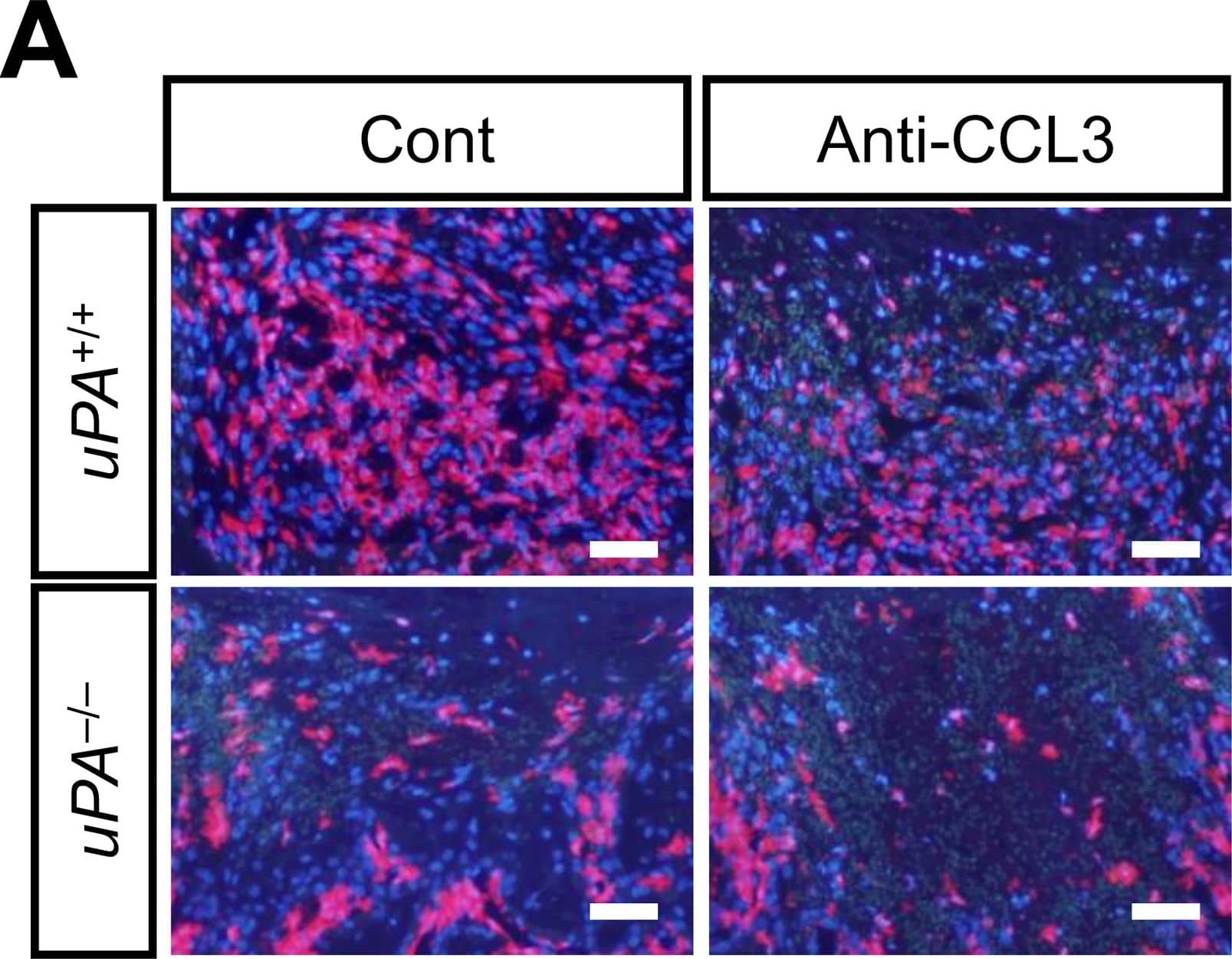
Detection of Mouse CCL3/MIP-1 alpha by Immunocytochemistry/ Immunofluorescence
Effects of neutralizing anti-CCL3 antibody on the number of macrophages at the damaged site and the decrease in area of bone defect on day 4 in uPA+/+ and uPA-/- mice.(A) Microphotographs of immunostaining for F4/80 at the damaged site on day 4 after a femoral bone defect in uPA+/+ and uPA-/- mice treated with normal IgG (Cont) or neutralizing anti-CCL3 antibody (Anti-CCL3). The results represent experiments performed on 5 mice in each group. Scale bars indicate 50 μm. (B) Quantification of the number of F4/80-positive cells per 0.1 mm2 in the microscopic fields in the damaged site on day 4 after a femoral bone defect in uPA+/+ and uPA-/- mice treated with normal IgG or neutralizing anti-CCL3 antibody. The data represent the mean ± SEM of 5 mice. (C) Quantification of the bone defect area as assessed by qCT on days 1 and 4 in uPA+/+ and uPA-/- mice treated with normal IgG or neutralizing anti-CCL3 antibody. The data represent the mean ± SEM from 5 mice. **P < 0.01 and *P < 0.05 (Tukey’s test). Image collected and cropped by CiteAb from the following open publication (https://dx.plos.org/10.1371/journal.pone.0123982), licensed under a CC-BY license. Not internally tested by R&D Systems.Applications for Mouse CCL3/MIP-1 alpha Antibody
Application
Recommended Usage
Western Blot
1 µg/mL
Sample: Recombinant Mouse CCL3/MIP-1 alpha Isoform LD78a (Catalog # 450-MA)
Sample: Recombinant Mouse CCL3/MIP-1 alpha Isoform LD78a (Catalog # 450-MA)
Neutralization
Measured by its ability to neutralize CCL3/MIP‑1 alpha-induced chemotaxis in BaF3 mouse pro‑B cell line transfected with human CCR5. The Neutralization Dose (ND50) is typically 0.3-1 µg/mL in the presence of 10 ng/mL Recombinant Mouse CCL3/MIP‑1 alpha Isoform LD78a.
Formulation, Preparation, and Storage
Purification
Protein A or G purified
Reconstitution
Reconstitute at 1 mg/mL in sterile PBS.
Formulation
Lyophilized from a 0.2 μm filtered solution in PBS with Trehalose.
Shipping
The product is shipped at ambient temperature. Upon receipt, store it immediately at the temperature recommended below.
Stability & Storage
Use a manual defrost freezer and avoid repeated freeze-thaw cycles.
- 12 months from date of receipt, -20 to -70 °C as supplied.
- 1 month, 2 to 8 °C under sterile conditions after reconstitution.
- 6 months, -20 to -70 °C under sterile conditions after reconstitution.
Background: CCL3/MIP-1 alpha
The macrophage inflammatory proteins 1 alpha and 1 beta, two closely related but distinct proteins, were originally co-purified from medium conditioned by a LPS-stimulated murine macrophage cell line. Mature mouse MIP-1 alpha shares approximately 77% and 70% amino acid identity with human MIP-1 alpha and mouse MIP-1 beta, respectively.
MIP‑1 proteins are expressed primarily in T cells, B cells, and monocytes after antigen or mitogen stimulation. The MIP-1 proteins are members of the beta (C-C) subfamily of chemokines.
Both MIP-1 alpha and MIP-1 beta are monocyte chemoattractants in vitro. Additionally, the MIP-1 proteins have been reported to have chemoattractant and adhesive effects on lymphocytes, with MIP-1 alpha and MIP-1 beta preferentially attracting CD8+ and CD4+ T cells, respectively. MIP-1 alpha has also been shown to attract B cells as well as eosinophils. MIP-1 proteins have been reported to have multiple effects on hematopoietic precursor cells and MIP-1 alpha has been identified as a stem cell inhibitory factor that can inhibit the proliferation of hematopoietic stem cells in vitro as well as in vivo. In the same assays, MIP-1 beta was reported to be much less active. The functional receptor for MIP-1 alpha has been identified as CCR1 and CCR5.
References
- Menten, P. et al. (2002) Cytokine Growth Factor Rev. 13:455.
Alternate Names
G0S19-1, LD78a, MIP-1 alpha, MIP1 alpha, MIP1A, PAT 464.1, SCYA3, SIS-beta
Entrez Gene IDs
Gene Symbol
CCL3
UniProt
Additional CCL3/MIP-1 alpha Products
Product Documents for Mouse CCL3/MIP-1 alpha Antibody
Product Specific Notices for Mouse CCL3/MIP-1 alpha Antibody
For research use only
Loading...
Loading...
Loading...
Loading...
Loading...